9TLJ | pdb_00009tlj

Horse hemoglobin structure from serial crystallography with 10 fs pulse


Experimental Data Snapshot

  • Method: X-RAY DIFFRACTION
  • Resolution: 2.46 Å
  • R-Value Free: 
    0.227 (Depositor), 0.233 (DCC) 
  • R-Value Work: 
    0.172 (Depositor), 0.180 (DCC) 
  • R-Value Observed: 
    0.175 (Depositor) 

Starting Model: experimental
View more details

wwPDB Validation 3D Report Full Report

Validation slider image for 9TLJ

Ligand Structure Quality Assessment 


This is version 1.0 of the entry. See complete history. 

Literature

Radiation damage in serial femtosecond crystallography studied in hemoglobin.

Galchenkova, M., Dawod, I., Cardoch, S., Sprenger, J., Oberthur, D., Awel, S., De Santis, E., Granas, O., Knoska, J., Metz, M., Passmore, S., Pena, G., Sarrou, I., Wiedorn, M., Timneanu, N., Chapman, H., Yefanov, O., Caleman, C.

(2026) Acta Crystallogr D Struct Biol 82: 1169-1180

  • DOI: https://doi.org/10.1107/S2059798326008569
  • Primary Citation Related Structures: 
    9TLI, 9TLJ

  • PubMed Abstract: 

    A central question in macromolecular crystallography is what X-ray free-electron laser pulse duration is required to obtain damage-free structural information. We compare serial femtosecond crystallography data from hemoglobin crystals using 3 and 10 fs pulses at comparable peak intensities (∼3 × 10 17  W cm -2 ) and fixed photon energy (7.15 keV). Structural refinement produced very similar models and resolution-dependent data-quality indicators remained comparable. Both datasets also retained sufficient anomalous signal for phasing. These observations support the conclusion that high-resolution scattering was preserved under both pulse durations. Hybrid collisional-radiative and molecular-dynamics simulations show that under typical experimental conditions, atomic form-factor changes are negligible (<1%) and atomic displacements fall below the resolution limit imposed by the X-ray photon energy and the geometry of the detector. The combined experimental and theoretical results indicate that 10 fs pulses are adequate to obtain damage-free protein structures using femtosecond crystallography under the intensity conditions explored.


  • Organizational Affiliation: 
    • Center for Free-Electron Laser Science (CFEL), Deutsches Elektronen-Synchrotron DESY, Notkestrasse 85, 22607 Hamburg, Germany.

Macromolecule Content 

  • Total Structure Weight: 65.26 kDa 
  • Atom Count: 4,828 
  • Modeled Residue Count: 572 
  • Deposited Residue Count: 576 
  • Unique protein chains: 2

Macromolecules

Find similar proteins by:|  3D Structure
Entity ID: 1
MoleculeChains  Sequence LengthOrganismDetailsImage
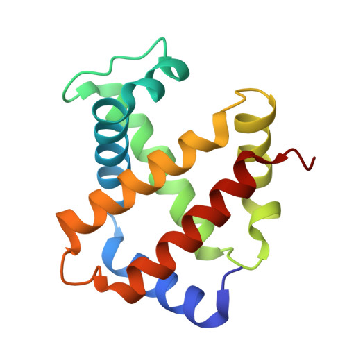
Hemoglobin subunit alpha
A, C
142Equus caballusMutation(s): 0 
UniProt
Find proteins for P01958 (Equus caballus)
Explore P01958 
Go to UniProtKB:  P01958
Entity Groups
Sequence Clusters30% Identity50% Identity70% Identity90% Identity95% Identity100% Identity
UniProt GroupP01958
Sequence Annotations
Expand
Reference Sequence
Find similar proteins by:|  3D Structure
Entity ID: 2
MoleculeChains  Sequence LengthOrganismDetailsImage
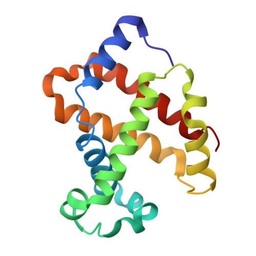
Hemoglobin subunit beta
B, D
146Equus caballusMutation(s): 0 
UniProt
Find proteins for P02062 (Equus caballus)
Explore P02062 
Go to UniProtKB:  P02062
Entity Groups
Sequence Clusters30% Identity50% Identity70% Identity90% Identity95% Identity100% Identity
UniProt GroupP02062
Sequence Annotations
Expand
Reference Sequence

Small Molecules

Ligands 4 Unique
IDChains Name / Formula / InChI Key2D Diagram3D Interactions
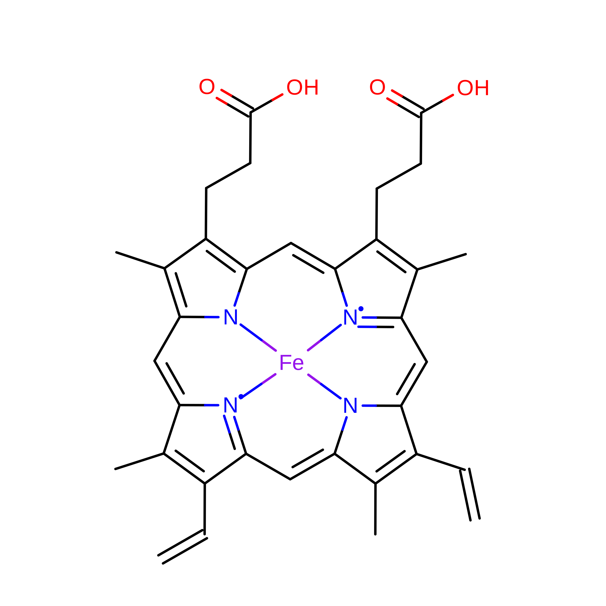
HEM
(Subject of Investigation/LOI)

Query on HEM


Download:Ideal Coordinates CCD File
E [auth A],
G [auth B],
H [auth C],
K [auth D]
PROTOPORPHYRIN IX CONTAINING FE
C34 H32 Fe N4 O4
KABFMIBPWCXCRK-RGGAHWMASA-L
PEG

Query on PEG


Download:Ideal Coordinates CCD File
L [auth D]DI(HYDROXYETHYL)ETHER
C4 H10 O3
MTHSVFCYNBDYFN-UHFFFAOYSA-N
CL

Query on CL


Download:Ideal Coordinates CCD File
J [auth C]CHLORIDE ION
Cl
VEXZGXHMUGYJMC-UHFFFAOYSA-M
NA

Query on NA


Download:Ideal Coordinates CCD File
F [auth A],
I [auth C]
SODIUM ION
Na
FKNQFGJONOIPTF-UHFFFAOYSA-N

Experimental Data & Validation

Experimental Data

  • Method: X-RAY DIFFRACTION
  • Resolution: 2.46 Å
  • R-Value Free:  0.227 (Depositor), 0.233 (DCC) 
  • R-Value Work:  0.172 (Depositor), 0.180 (DCC) 
  • R-Value Observed: 0.175 (Depositor) 
Space Group: P 21 21 21
Unit Cell:
Length ( Å )Angle ( ˚ )
a = 62.51α = 90
b = 80.97β = 90
c = 111.48γ = 90
Software Package:
Software NamePurpose
PHENIXrefinement
PDB_EXTRACTdata extraction
CrystFELdata reduction
CrystFELdata scaling
PHASERphasing

Structure Validation

View Full Validation Report


Ligand Structure Quality Assessment 


Entry History 

& Funding Information

Deposition Data


Funding OrganizationLocationGrant Number
Swedish Research CouncilSweden018-00740, 2019-0393

Revision History  (Full details and data files)

  • Version 1.0: 2026-10-14
    Type: Initial release