Download Mendeley
Radiation damage in serial femtosecond crystallography studied in hemoglobin.
Galchenkova, M., Dawod, I., Cardoch, S., Sprenger, J., Oberthur, D., Awel, S., De Santis, E., Granas, O., Knoska, J., Metz, M., Passmore, S., Pena, G., Sarrou, I., Wiedorn, M., Timneanu, N., Chapman, H., Yefanov, O., Caleman, C.(2026) Acta Crystallogr D Struct Biol 82: 1169-1180
- PubMed: 42813594 Search on PubMedSearch on PubMed Central
- DOI: https://doi.org/10.1107/S2059798326008569
- Primary Citation Related Structures:
9TLI, 9TLJ - PubMed Abstract:
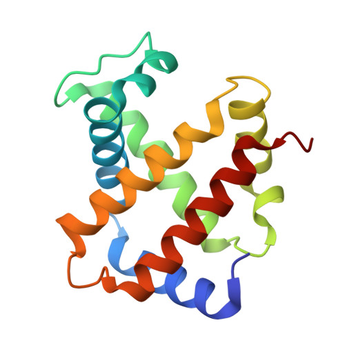
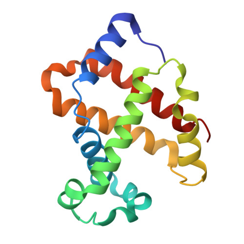
A central question in macromolecular crystallography is what X-ray free-electron laser pulse duration is required to obtain damage-free structural information. We compare serial femtosecond crystallography data from hemoglobin crystals using 3 and 10 fs pulses at comparable peak intensities (∼3 × 10 17 W cm -2 ) and fixed photon energy (7.15 keV). Structural refinement produced very similar models and resolution-dependent data-quality indicators remained comparable. Both datasets also retained sufficient anomalous signal for phasing. These observations support the conclusion that high-resolution scattering was preserved under both pulse durations. Hybrid collisional-radiative and molecular-dynamics simulations show that under typical experimental conditions, atomic form-factor changes are negligible (<1%) and atomic displacements fall below the resolution limit imposed by the X-ray photon energy and the geometry of the detector. The combined experimental and theoretical results indicate that 10 fs pulses are adequate to obtain damage-free protein structures using femtosecond crystallography under the intensity conditions explored.
- Center for Free-Electron Laser Science (CFEL), Deutsches Elektronen-Synchrotron DESY, Notkestrasse 85, 22607 Hamburg, Germany.
Organizational Affiliation: