9R3M | pdb_00009r3m

Structure of liver pyruvate kinase in complex with fluorescent probe 8a


Experimental Data Snapshot

  • Method: X-RAY DIFFRACTION
  • Resolution: 2.06 Å
  • R-Value Free: 
    0.235 (Depositor), 0.224 (DCC) 
  • R-Value Work: 
    0.199 (Depositor), 0.190 (DCC) 
  • R-Value Observed: 
    0.201 (Depositor) 

Starting Model: experimental
View more details

wwPDB Validation   3D Report Full Report


Ligand Structure Quality Assessment 


This is version 1.0 of the entry. See complete history


Literature

Fluorescent binding assay for allosteric ligands of liver pyruvate kinase.

Nilsson, O.Bogucka, A.Koteles, I.Haversen, L.Liljenberg, S.Rutberg, M.Hyvonen, M.Grotli, M.

(2025) Eur J Med Chem 298: 117989-117989

  • DOI: https://doi.org/10.1016/j.ejmech.2025.117989
  • Primary Citation of Related Structures:  
    9R3H, 9R3I, 9R3L, 9R3M, 9R3O

  • PubMed Abstract: 

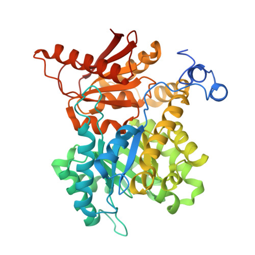
    Environment-sensitive fluorescent probes are indispensable tools for studying biological systems and advancing drug discovery. This study reports the development of 4-sulfamoyl-7-aminobenzoxadiazole (SBD)-based fluorescent probes for the allosteric site of the liver isoform of pyruvate kinase (PKL). By integrating SBD moieties into known activator scaffolds, such as mitapivat and diarylsulfonamide (DASA) ligands, probes for indicator displacement assays were designed to quantify ligand interactions in the allosteric site. Compound 4a displayed dose-dependent fluorescence enhancement in response to PKL binding and was used in a competitive binding assay with unlabelled ligands: mitapivat, TEPP-46, DASA-58 and reported activator 21. Structure-activity relationship (SAR) analysis revealed key structural features influencing activity and fluorescence sensitivity. The probes report selectively on the allosteric site ligands as the binding was not affected by natural ligands, such as ADP, fructose-1,6-bisphosphate (FBP), phosphoenolpyruvate (PEP), and phenylalanine. These findings provide a practical framework for detecting allosteric ligand engagement in PKL and expand the repertoire of molecular tools for advancing PKL-targeted therapies.


  • Organizational Affiliation

    Department of Chemistry and Molecular Biology, University of Gothenburg, Medicinaregatan 7B, 413 90, Gothenburg, Sweden.


Macromolecules
Find similar proteins by:  (by identity cutoff)  |  3D Structure
Entity ID: 1
MoleculeChains Sequence LengthOrganismDetailsImage
Isoform L-type of Pyruvate kinase PKLR
A, B, C, D, E
A, B, C, D, E, F, G, H
447Homo sapiensMutation(s): 0 
Gene Names: PKLRPK1PKL
EC: 2.7.1.40
UniProt & NIH Common Fund Data Resources
Find proteins for P30613 (Homo sapiens)
Go to UniProtKB:  P30613
PHAROS:  P30613
GTEx:  ENSG00000143627 
Entity Groups  
Sequence Clusters30% Identity50% Identity70% Identity90% Identity95% Identity100% Identity
UniProt GroupP30613-2
Sequence Annotations
Expand
  • Reference Sequence
Small Molecules
Ligands 4 Unique
IDChains Name / Formula / InChI Key2D Diagram3D Interactions
FBP
Query on FBP

Download Ideal Coordinates CCD File 
AA [auth E]
FA [auth F]
I [auth A]
KA [auth G]
N [auth B]
AA [auth E],
FA [auth F],
I [auth A],
KA [auth G],
N [auth B],
OA [auth H],
R [auth C],
V [auth D]
1,6-di-O-phosphono-beta-D-fructofuranose
C6 H14 O12 P2
RNBGYGVWRKECFJ-ARQDHWQXSA-N
OXL
Query on OXL

Download Ideal Coordinates CCD File 
BA [auth E]
GA [auth F]
J [auth A]
LA [auth G]
O [auth B]
BA [auth E],
GA [auth F],
J [auth A],
LA [auth G],
O [auth B],
PA [auth H],
S [auth C],
W [auth D]
OXALATE ION
C2 O4
MUBZPKHOEPUJKR-UHFFFAOYSA-L
K
Query on K

Download Ideal Coordinates CCD File 
DA [auth E]
IA [auth F]
L [auth A]
NA [auth G]
Q [auth B]
DA [auth E],
IA [auth F],
L [auth A],
NA [auth G],
Q [auth B],
RA [auth H],
U [auth C],
Y [auth D]
POTASSIUM ION
K
NPYPAHLBTDXSSS-UHFFFAOYSA-N
MG
Query on MG

Download Ideal Coordinates CCD File 
CA [auth E]
HA [auth F]
K [auth A]
MA [auth G]
P [auth B]
CA [auth E],
HA [auth F],
K [auth A],
MA [auth G],
P [auth B],
QA [auth H],
T [auth C],
X [auth D]
MAGNESIUM ION
Mg
JLVVSXFLKOJNIY-UHFFFAOYSA-N
Experimental Data & Validation

Experimental Data

  • Method: X-RAY DIFFRACTION
  • Resolution: 2.06 Å
  • R-Value Free:  0.235 (Depositor), 0.224 (DCC) 
  • R-Value Work:  0.199 (Depositor), 0.190 (DCC) 
  • R-Value Observed: 0.201 (Depositor) 
Space Group: C 1 2 1
Unit Cell:
Length ( Å )Angle ( ˚ )
a = 208.443α = 90
b = 112.896β = 91.06
c = 189.009γ = 90
Software Package:
Software NamePurpose
autoPROCdata processing
XDSdata reduction
Aimlessdata scaling
STARANISOdata scaling
BUSTERrefinement
PHASERphasing

Structure Validation

View Full Validation Report


Ligand Structure Quality Assessment 


Entry History & Funding Information

Deposition Data


Funding OrganizationLocationGrant Number
Wellcome TrustUnited Kingdom223804/Z/21/Z

Revision History  (Full details and data files)

  • Version 1.0: 2025-08-13
    Type: Initial release