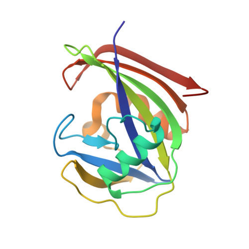
9MBL | pdb_00009mbl

2-Oxo-dATP hydrolysis in human MTH1(G2K mutant) crystal using Mn2+: the ES-2M complex


Experimental Data Snapshot

  • Method: X-RAY DIFFRACTION
  • Resolution: 1.37 Å
  • R-Value Free: 
    0.181 (Depositor), 0.180 (DCC) 
  • R-Value Work: 
    0.134 (Depositor), 0.134 (DCC) 
  • R-Value Observed: 
    0.136 (Depositor) 

Starting Model: experimental
View more details

wwPDB Validation   3D Report Full Report


Ligand Structure Quality Assessment 


This is version 1.0 of the entry. See complete history


Literature

Neutron and time-resolved X-ray crystallography reveal the substrate recognition and catalytic mechanism of human Nudix hydrolase MTH1.

Hirata, K.Fujimiya, K.Ostermann, A.Schrader, T.E.Hiromoto, T.Goto, M.Arimori, T.Hirano, Y.Kusaka, K.Tamada, T.Nakamura, T.

(2025) Proc Natl Acad Sci U S A 122: e2510085122-e2510085122

  • DOI: https://doi.org/10.1073/pnas.2510085122
  • Primary Citation of Related Structures:  
    9MBE, 9MBF, 9MBG, 9MBH, 9MBI, 9MBJ, 9MBK, 9MBL, 9MBM, 9MBN

  • PubMed Abstract: 

    Human MTH1, a Nudix enzyme, hydrolyzes several oxidized nucleotides such as 8-oxo-dGTP and 2-oxo-dATP, owing to its broad substrate specificity. MTH1 has also attracted attention as an anticancer target, and its substrate recognition is of biological and medical interest. Previous studies have suggested that MTH1 exhibits broad substrate specificity by changing the protonation states of Asp119 and Asp120 with high p K a . However, the recognition mechanism remains unclear, owing to the difficulty in directly observing hydrogen atoms. Furthermore, a recent time-resolved X-ray study has proposed that Nudix hydrolases catalyze reactions through a new three-metal-ion mechanism. To understand the substrate recognition and catalytic mechanisms of human MTH1, we performed neutron and time-resolved X-ray crystallography. The neutron structures of MTH1 complexed with 8-oxo-dGTP and 2-oxo-dATP revealed the protonation states of the active-site residues, substrates, and water molecules, crucial for substrate binding and catalysis, providing direct experimental evidence that changes in the protonation states of Asp119 and Asp120 enable broad substrate recognition of MTH1. Time-resolved X-ray crystallography was used to visualize the entire reaction process through Mn 2+ ion. The combination of neutron and time-resolved X-ray crystallography led to the proposal of a reaction mechanism for MTH1 via three metal-binding sites, including the conformational dynamics of a loop region, nucleophilic substitution, and a potential deprotonation pathway. Overall, the mechanism involving three metal-binding sites may be a general feature in the catalysis of Nudix hydrolases.


  • Organizational Affiliation

    Graduate School of Pharmaceutical Sciences, Kumamoto University, Kumamoto 862-0973, Japan.


Macromolecules
Find similar proteins by:  (by identity cutoff)  |  3D Structure
Entity ID: 1
MoleculeChains Sequence LengthOrganismDetailsImage
7,8-dihydro-8-oxoguanine triphosphatase
A, B
156Homo sapiensMutation(s): 1 
Gene Names: NUDT1MTH1
EC: 3.6.1.55 (PDB Primary Data), 3.6.1.56 (PDB Primary Data), 3.6.1 (UniProt)
UniProt & NIH Common Fund Data Resources
Find proteins for P36639 (Homo sapiens)
Explore P36639 
Go to UniProtKB:  P36639
PHAROS:  P36639
GTEx:  ENSG00000106268 
Entity Groups  
Sequence Clusters30% Identity50% Identity70% Identity90% Identity95% Identity100% Identity
UniProt GroupP36639
Sequence Annotations
Expand
  • Reference Sequence
Small Molecules
Ligands 3 Unique
IDChains Name / Formula / InChI Key2D Diagram3D Interactions
6U4 (Subject of Investigation/LOI)
Query on 6U4

Download Ideal Coordinates CCD File 
C [auth A],
H [auth B]
[[(2R,3S,5R)-5-(6-azanyl-2-oxidanylidene-1H-purin-9-yl)-3-oxidanyl-oxolan-2-yl]methoxy-oxidanyl-phosphoryl] phosphono hydrogen phosphate
C10 H16 N5 O13 P3
UOACBPRDWRDEHJ-KVQBGUIXSA-N
MN (Subject of Investigation/LOI)
Query on MN

Download Ideal Coordinates CCD File 
D [auth A],
E [auth A],
I [auth B],
J [auth B]
MANGANESE (II) ION
Mn
WAEMQWOKJMHJLA-UHFFFAOYSA-N
NA (Subject of Investigation/LOI)
Query on NA

Download Ideal Coordinates CCD File 
F [auth A],
G [auth A],
K [auth B],
L [auth B]
SODIUM ION
Na
FKNQFGJONOIPTF-UHFFFAOYSA-N
Experimental Data & Validation

Experimental Data

  • Method: X-RAY DIFFRACTION
  • Resolution: 1.37 Å
  • R-Value Free:  0.181 (Depositor), 0.180 (DCC) 
  • R-Value Work:  0.134 (Depositor), 0.134 (DCC) 
  • R-Value Observed: 0.136 (Depositor) 
Space Group: P 21 21 21
Unit Cell:
Length ( Å )Angle ( ˚ )
a = 46.503α = 90
b = 47.357β = 90
c = 123.631γ = 90
Software Package:
Software NamePurpose
PHENIXrefinement
HKL-2000data reduction
HKL-2000data scaling
PHENIXphasing

Structure Validation

View Full Validation Report


Ligand Structure Quality Assessment 


Entry History & Funding Information

Deposition Data


Funding OrganizationLocationGrant Number
Japan Society for the Promotion of Science (JSPS)Japan--

Revision History  (Full details and data files)

  • Version 1.0: 2025-07-30
    Type: Initial release