Neutron and time-resolved X-ray crystallography reveal the substrate recognition and catalytic mechanism of human Nudix hydrolase MTH1.
Hirata, K., Fujimiya, K., Ostermann, A., Schrader, T.E., Hiromoto, T., Goto, M., Arimori, T., Hirano, Y., Kusaka, K., Tamada, T., Nakamura, T.(2025) Proc Natl Acad Sci U S A 122: e2510085122-e2510085122
- PubMed: 40674425
- DOI: https://doi.org/10.1073/pnas.2510085122
- Primary Citation of Related Structures:
9MBE, 9MBF, 9MBG, 9MBH, 9MBI, 9MBJ, 9MBK, 9MBL, 9MBM, 9MBN - PubMed Abstract:
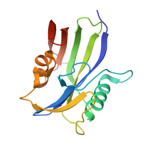
Human MTH1, a Nudix enzyme, hydrolyzes several oxidized nucleotides such as 8-oxo-dGTP and 2-oxo-dATP, owing to its broad substrate specificity. MTH1 has also attracted attention as an anticancer target, and its substrate recognition is of biological and medical interest. Previous studies have suggested that MTH1 exhibits broad substrate specificity by changing the protonation states of Asp119 and Asp120 with high p K a . However, the recognition mechanism remains unclear, owing to the difficulty in directly observing hydrogen atoms. Furthermore, a recent time-resolved X-ray study has proposed that Nudix hydrolases catalyze reactions through a new three-metal-ion mechanism. To understand the substrate recognition and catalytic mechanisms of human MTH1, we performed neutron and time-resolved X-ray crystallography. The neutron structures of MTH1 complexed with 8-oxo-dGTP and 2-oxo-dATP revealed the protonation states of the active-site residues, substrates, and water molecules, crucial for substrate binding and catalysis, providing direct experimental evidence that changes in the protonation states of Asp119 and Asp120 enable broad substrate recognition of MTH1. Time-resolved X-ray crystallography was used to visualize the entire reaction process through Mn 2+ ion. The combination of neutron and time-resolved X-ray crystallography led to the proposal of a reaction mechanism for MTH1 via three metal-binding sites, including the conformational dynamics of a loop region, nucleophilic substitution, and a potential deprotonation pathway. Overall, the mechanism involving three metal-binding sites may be a general feature in the catalysis of Nudix hydrolases.
- Graduate School of Pharmaceutical Sciences, Kumamoto University, Kumamoto 862-0973, Japan.
Organizational Affiliation: