9O3B | pdb_00009o3b

PKM2 bound to MCTI-566

  • Classification: TRANSFERASE
  • Organism(s): Homo sapiens
  • Expression System: Escherichia coli
  • Mutation(s): No 

  • Deposited: 2025-04-07 Released: 2025-08-13 
  • Deposition Author(s): Stuckey, J.A.
  • Funding Organization(s): National Institutes of Health/National Eye Institute (NIH/NEI)

Experimental Data Snapshot

  • Method: X-RAY DIFFRACTION
  • Resolution: 2.42 Å
  • R-Value Free: 
    0.226 (Depositor), 0.223 (DCC) 
  • R-Value Work: 
    0.186 (Depositor), 0.180 (DCC) 
  • R-Value Observed: 
    0.188 (Depositor) 

Starting Model: experimental
View more details

wwPDB Validation   3D Report Full Report


Ligand Structure Quality Assessment 


This is version 1.0 of the entry. See complete history


Literature

A Novel, Long-Acting, Small Molecule PKM2 Activator and Its Potential Broad Application Against Photoreceptor Degeneration.

Pan, W.W.Weh, K.M.Chaudhury, S.Fernando, R.Hager, H.Wen, B.Chinnaswamy, K.Stuckey, J.A.Rech, J.C.Besirli, C.G.Weh, E.Wubben, T.J.

(2025) Transl Vis Sci Technol 14: 26-26

  • DOI: https://doi.org/10.1167/tvst.14.7.26
  • Primary Citation of Related Structures:  
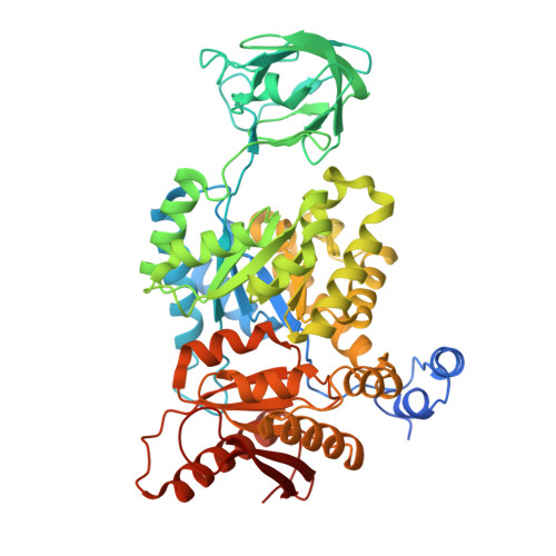
    9O3B

  • PubMed Abstract: 

    Activating pyruvate kinase M2 (PKM2) has been shown to be neuroprotective in preclinical models of photoreceptor degeneration. We recently developed novel, small molecule activators for ocular delivery. Here, we sought to characterize the ocular pharmacology, toxicity, and efficacy of MCTI-566, a novel PKM2 activator, to translate this therapeutic strategy to the clinic. X-ray protein crystallography and isothermal titration calorimetry assessed the interaction of MCTI-566 with PKM2. PKM2 activation and tissue pharmacokinetics were examined after intravitreal or systemic administration of MCTI-566. Retinal toxicity was evaluated in rats after intravitreal injection. The effect of MCTI-566 on photoreceptor death was assessed using in vitro and in vivo models of outer retinal stress and on the inflammatory response in the rd10 retina using flow cytometry and quantitative real-time polymerase chain reaction. The PKM2-MCTI-566 co-crystal structure demonstrated a binding pocket distinct from endogenous activators. MCTI-566 increases retinal PK activity 200% following intravitreal or systemic administration. MCTI-566 distributed to the retina after intravitreal or systemic administration, activated the target for ≥90 days and was specific for photoreceptor PKM2. No retinal toxicity was observed after repeated intravitreal administration. MCTI-566 reduced photoreceptor apoptosis in a model of retinal detachment, and delayed photoreceptor degeneration and altered the inflammatory response in the rd10 retina. MCTI-566 is a small molecule drug candidate for photoreceptor neuroprotection. MCTI-566, a long-acting and well-tolerated ocular PKM2 activator, may be a potential therapeutic to combat currently untreatable retinal degenerations.


  • Organizational Affiliation

    Department of Ophthalmology and Visual Sciences, Kellogg Eye Center, University of Michigan, Ann Arbor, MI, USA.


Macromolecules
Find similar proteins by:  (by identity cutoff)  |  3D Structure
Entity ID: 1
MoleculeChains Sequence LengthOrganismDetailsImage
Pyruvate kinase PKM
A, B, C, D, E
A, B, C, D, E, F, G, H
550Homo sapiensMutation(s): 0 
Gene Names: PKMOIP3PK2PK3PKM2
EC: 2.7.1.40 (PDB Primary Data), 2.7.11.1 (UniProt), 2.7.10.2 (UniProt)
UniProt & NIH Common Fund Data Resources
Find proteins for P14618 (Homo sapiens)
Explore P14618 
Go to UniProtKB:  P14618
PHAROS:  P14618
GTEx:  ENSG00000067225 
Entity Groups  
Sequence Clusters30% Identity50% Identity70% Identity90% Identity95% Identity100% Identity
UniProt GroupP14618
Sequence Annotations
Expand
  • Reference Sequence
Small Molecules
Ligands 5 Unique
IDChains Name / Formula / InChI Key2D Diagram3D Interactions
FBP
Query on FBP

Download Ideal Coordinates CCD File 
BA [auth F],
I [auth A],
N [auth B],
X [auth E]
1,6-di-O-phosphono-beta-D-fructofuranose
C6 H14 O12 P2
RNBGYGVWRKECFJ-ARQDHWQXSA-N
PO4
Query on PO4

Download Ideal Coordinates CCD File 
GA [auth G],
LA [auth H],
U [auth C],
W [auth D]
PHOSPHATE ION
O4 P
NBIIXXVUZAFLBC-UHFFFAOYSA-K
OXL
Query on OXL

Download Ideal Coordinates CCD File 
CA [auth F],
HA [auth H],
J [auth A],
Q [auth C]
OXALATE ION
C2 O4
MUBZPKHOEPUJKR-UHFFFAOYSA-L
EDO
Query on EDO

Download Ideal Coordinates CCD File 
EA [auth G]
JA [auth H]
K [auth A]
O [auth B]
S [auth C]
EA [auth G],
JA [auth H],
K [auth A],
O [auth B],
S [auth C],
Y [auth E]
1,2-ETHANEDIOL
C2 H6 O2
LYCAIKOWRPUZTN-UHFFFAOYSA-N
MG
Query on MG

Download Ideal Coordinates CCD File 
DA [auth F]
FA [auth G]
KA [auth H]
L [auth A]
P [auth B]
DA [auth F],
FA [auth G],
KA [auth H],
L [auth A],
P [auth B],
T [auth C],
V [auth D],
Z [auth E]
MAGNESIUM ION
Mg
JLVVSXFLKOJNIY-UHFFFAOYSA-N
Experimental Data & Validation

Experimental Data

  • Method: X-RAY DIFFRACTION
  • Resolution: 2.42 Å
  • R-Value Free:  0.226 (Depositor), 0.223 (DCC) 
  • R-Value Work:  0.186 (Depositor), 0.180 (DCC) 
  • R-Value Observed: 0.188 (Depositor) 
Space Group: P 1
Unit Cell:
Length ( Å )Angle ( ˚ )
a = 80.807α = 89.73
b = 93.855β = 89.68
c = 153.66γ = 75.79
Software Package:
Software NamePurpose
BUSTERrefinement
HKL-2000data scaling
HKL-2000data reduction
MOLREPphasing
PDB_EXTRACTdata extraction

Structure Validation

View Full Validation Report


Ligand Structure Quality Assessment 


Entry History & Funding Information

Deposition Data


Funding OrganizationLocationGrant Number
National Institutes of Health/National Eye Institute (NIH/NEI)United StatesK08EY031757

Revision History  (Full details and data files)

  • Version 1.0: 2025-08-13
    Type: Initial release