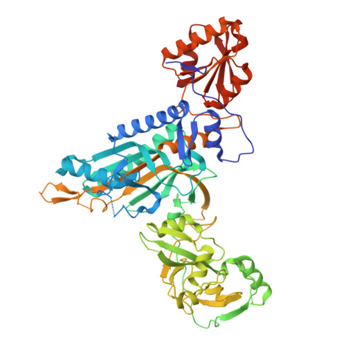
9Z09 | pdb_00009z09

Crystal structure of M. tuberculosis ProRS bound to 3-(4-bromobenzamido)pyrazine-2-carboxamide (compound 4e)


Experimental Data Snapshot

  • Method: X-RAY DIFFRACTION
  • Resolution: 2.60 Å
  • R-Value Free: 
    0.258 (Depositor), 0.258 (DCC) 
  • R-Value Work: 
    0.214 (Depositor), 0.214 (DCC) 
  • R-Value Observed: 
    0.216 (Depositor) 

Starting Model: in silico
View more details

wwPDB Validation 3D Report Full Report

Validation slider image for 9Z09

Ligand Structure Quality Assessment 


This is version 1.0 of the entry. See complete history. 

Literature

Atomic-Level Structures Reveal How a Repurposed 3‐Aminopyrazine-2-carboxamide Scaffold Inhibits Mycobacterium tuberculosis Prolyl-tRNA Synthetase.

Gade, P., Michalska, K., Zitko, J., Wower, J., Maltseva, N., Pallabothula, V.S.K., Jedrzejczak, R., Joachimiak, A.

(2026) ACS Omega 11: 57583-57594

  • DOI: https://doi.org/10.1021/acsomega.6c04844
  • Primary Citation Related Structures: 
    9YXS, 9YY6, 9YYE, 9YYF, 9YYO, 9YYT, 9YZX, 9Z09

  • PubMed Abstract: 

    Tuberculosis (TB) is again the world's leading cause of death from a single infectious agent. Aminoacyl-tRNA synthetases are essential for protein synthesis and are promising drug targets because bacterial and human enzymes differ. We repurposed a human prolyl-tRNA synthetase (ProRS) inhibitor scaffold, 3-aminopyrazine-2-carboxamide, to inhibit the Mycobacterium tuberculosis enzyme ( Mtb ProRS). These derivatives show strong activity against multidrug-resistant Mtb strains and are not cytotoxic to HepG2 cells. We determined high-resolution crystal structures of Mtb ProRS bound to six derivatives to define their binding modes. The compounds bind in the ATP site and trigger local conformational changes that disrupt the proline pocket, supporting a dual-site mechanism of inhibition. Comparison with human cytosolic and mitochondrial ProRS reveals differences in active-site residues, electrostatics, and dynamics that explain species selectivity. Together, these structures provide a framework for designing next-generation, Mtb -selective ProRS inhibitors as new antitubercular candidates.


  • Organizational Affiliation: 
    • Center for Structural Biology of Infectious Diseases, Consortium for Advanced Science and Engineering, University of Chicago, Chicago, Illinois 60667, United States.

Macromolecule Content 

  • Total Structure Weight: 129.36 kDa 
  • Atom Count: 8,749 
  • Modeled Residue Count: 1,122 
  • Deposited Residue Count: 1,176 
  • Unique protein chains: 1

Macromolecules

Find similar proteins by:|  3D Structure
Entity ID: 1
MoleculeChains  Sequence LengthOrganismDetailsImage
Proline--tRNA ligase
A, B
588Mycobacterium tuberculosis H37RvMutation(s): 0 
Gene Names: proS, Rv2845c, MTCY24A1.12
EC: 6.1.1.15
UniProt
Find proteins for P9WFT9 (Mycobacterium tuberculosis (strain ATCC 25618 / H37Rv))
Explore P9WFT9 
Go to UniProtKB:  P9WFT9
Entity Groups
Sequence Clusters30% Identity50% Identity70% Identity90% Identity95% Identity100% Identity
UniProt GroupP9WFT9
Sequence Annotations
Expand
Reference Sequence

Small Molecules

Ligands 5 Unique
IDChains Name / Formula / InChI Key2D Diagram3D Interactions
A1CZT
(Subject of Investigation/LOI)

Query on A1CZT


Download:Ideal Coordinates CCD File
C [auth A],
J [auth B]
3-(4-bromobenzamido)pyrazine-2-carboxamide
C12 H9 Br N4 O2
SAZAWTYKBYDDJF-UHFFFAOYSA-N
EDO

Query on EDO


Download:Ideal Coordinates CCD File
F [auth A],
G [auth A],
I [auth A]
1,2-ETHANEDIOL
C2 H6 O2
LYCAIKOWRPUZTN-UHFFFAOYSA-N
ACT

Query on ACT


Download:Ideal Coordinates CCD File
H [auth A]ACETATE ION
C2 H3 O2
QTBSBXVTEAMEQO-UHFFFAOYSA-M
CA

Query on CA


Download:Ideal Coordinates CCD File
E [auth A],
L [auth B]
CALCIUM ION
Ca
BHPQYMZQTOCNFJ-UHFFFAOYSA-N
MG

Query on MG


Download:Ideal Coordinates CCD File
D [auth A],
K [auth B]
MAGNESIUM ION
Mg
JLVVSXFLKOJNIY-UHFFFAOYSA-N

Experimental Data & Validation

Experimental Data

  • Method: X-RAY DIFFRACTION
  • Resolution: 2.60 Å
  • R-Value Free:  0.258 (Depositor), 0.258 (DCC) 
  • R-Value Work:  0.214 (Depositor), 0.214 (DCC) 
  • R-Value Observed: 0.216 (Depositor) 
Space Group: P 1 2 1
Unit Cell:
Length ( Å )Angle ( ˚ )
a = 85.874α = 90
b = 59.598β = 91.04
c = 136.053γ = 90
Software Package:
Software NamePurpose
PHENIXrefinement
Aimlessdata scaling
autoPROCdata reduction
MOLREPphasing
PDB_EXTRACTdata extraction

Structure Validation

View Full Validation Report


Ligand Structure Quality Assessment 


Entry History 

& Funding Information

Deposition Data


Funding OrganizationLocationGrant Number
National Institutes of Health/National Institute Of Allergy and Infectious Diseases (NIH/NIAID)United States--

Revision History  (Full details and data files)

  • Version 1.0: 2026-11-04
    Type: Initial release