9Y0E | pdb_00009y0e

Crystal structure of Bet v 1.0101 in complex with human IgE Fab 2H22


Experimental Data Snapshot

  • Method: X-RAY DIFFRACTION
  • Resolution: 2.90 Å
  • R-Value Free: 
    0.257 (Depositor), 0.252 (DCC) 
  • R-Value Work: 
    0.196 (Depositor), 0.197 (DCC) 

Starting Model: experimental
View more details

wwPDB Validation   3D Report Full Report


Ligand Structure Quality Assessment 


This is version 1.0 of the entry. See complete history


Literature

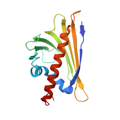
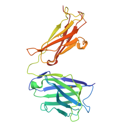
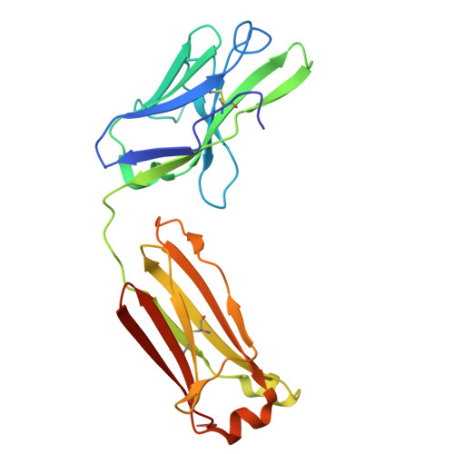
Structural Determination of a Human IgE Epitope on Major Birch Allergen Bet v 1.

O'Malley, A.Borders, B.T.Wilson, J.M.Smith, S.A.Chruszcz, M.

(2026) Allergy 

  • DOI: https://doi.org/10.1111/all.70240
  • Primary Citation of Related Structures:  
    9Y0A, 9Y0D, 9Y0E

  • Organizational Affiliation
    • Department of Biochemistry and Molecular Biology, Michigan State University, East Lansing, Michigan, USA.

Macromolecules
Find similar proteins by:  (by identity cutoff)  |  3D Structure
Entity ID: 1
MoleculeChains Sequence LengthOrganismDetailsImage
Major pollen allergen Bet v 1-AA [auth C]160Betula pendulaMutation(s): 0 
Gene Names: BETVIABETVI
UniProt
Find proteins for P15494 (Betula pendula)
Explore P15494 
Go to UniProtKB:  P15494
Entity Groups  
Sequence Clusters30% Identity50% Identity70% Identity90% Identity95% Identity100% Identity
UniProt GroupP15494
Sequence Annotations
Expand
  • Reference Sequence
Find similar proteins by:  (by identity cutoff)  |  3D Structure
Entity ID: 2
MoleculeChains Sequence LengthOrganismDetailsImage
hIgE Fab 2H22 heavy chainB [auth A]227Homo sapiensMutation(s): 0 
Entity Groups  
Sequence Clusters30% Identity50% Identity70% Identity90% Identity95% Identity100% Identity
Sequence Annotations
Expand
  • Reference Sequence
Find similar proteins by:  (by identity cutoff)  |  3D Structure
Entity ID: 3
MoleculeChains Sequence LengthOrganismDetailsImage
hIgE Fab 2H22 light chainC [auth B]216Homo sapiensMutation(s): 0 
Entity Groups  
Sequence Clusters30% Identity50% Identity70% Identity90% Identity95% Identity100% Identity
Glycosylation
Glycosylation Sites: 1
Sequence Annotations
Expand
  • Reference Sequence
Experimental Data & Validation

Experimental Data

  • Method: X-RAY DIFFRACTION
  • Resolution: 2.90 Å
  • R-Value Free:  0.257 (Depositor), 0.252 (DCC) 
  • R-Value Work:  0.196 (Depositor), 0.197 (DCC) 
Space Group: C 1 2 1
Unit Cell:
Length ( Å )Angle ( ˚ )
a = 150.971α = 90
b = 70.258β = 116.375
c = 78.247γ = 90
Software Package:
Software NamePurpose
REFMACrefinement
HKL-3000data reduction
HKL-3000data scaling
MOLREPphasing

Structure Validation

View Full Validation Report


Ligand Structure Quality Assessment 


Entry History & Funding Information

Deposition Data


Funding OrganizationLocationGrant Number
Other privateUnited States--

Revision History  (Full details and data files)

  • Version 1.0: 2026-02-11
    Type: Initial release