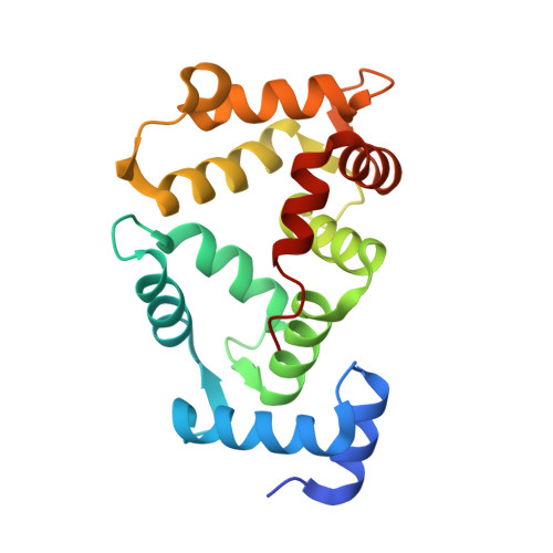
9QK7 | pdb_00009qk7

NCS-1 bound to XChem ligand 1


Experimental Data Snapshot

  • Method: X-RAY DIFFRACTION
  • Resolution: 2.15 Å
  • R-Value Free: 
    0.267 (Depositor), 0.262 (DCC) 
  • R-Value Work: 
    0.214 (Depositor), 0.215 (DCC) 
  • R-Value Observed: 
    0.217 (Depositor) 

Starting Model: experimental
View more details

wwPDB Validation 3D Report Full Report

Validation slider image for 9QK7

Ligand Structure Quality Assessment 


This is version 1.1 of the entry. See complete history. 

Literature

A resource-efficient structure-based workflow for fragment progression enables parallel hit discovery and validation of functionally diverse modulators of NCS-1 protein-protein interactions.

Munoz-Reyes, D., Fieseler, K.K., Winokan, M., Golding, M., Capkin, E., Ferla, M., Perez-Suarez, S., Tomlinson, C.W.E., Marples, P.G., Miro-Rodriguez, C., Aguado, L., Mansilla, A., Fearon, D., Thompson, W., von Delft, F., Sanchez-Barrena, M.J.

(2026) Chem Sci 

  • DOI: https://doi.org/10.1039/d6sc02031c
  • Primary Citation Related Structures: 
    9QK7, 9QK8, 9QK9, 9QKA, 9QKB, 9QKC, 9QKD, 9QKE, 9QKF, 9QKG, 9QKH, 9QKI, 9QKJ, 9QKK, 9QKL, 9QKM

  • PubMed Abstract: 

    Efficient drug discovery workflows ideally generate data that directly address key translational milestones, including confirmation of target engagement and binding pose, structure-activity relationships (SARs), and biological relevance, within rapid and resource-efficient experimental cycles. Here, we describe a data-driven, automation-assisted, customizable framework for fragment-to-hit progression that directly delivers structurally validated hit series primed for rapid SAR exploration by exploiting the high-throughput crystallography available at synchrotrons. This recently evolved direct-to-biology approach combines X-ray crystallographic fragment screening with algorithmically-guided fragment merging and reagent prioritization; low-cost robotic array synthesis and reaction production assessment by LC-MS; and finally orthogonal biophysical evaluation of crude reaction mixtures for binding assessment and 3D binding pose using grating-coupled interferometry and crystallography, respectively. We demonstrated the effectiveness of the strategy on a challenging target class by collectively progressing a large set of fragment hits through a single DMTA cycle comprising over 250 synthetically diverse compounds, enabling rapid, resource- and cost-effective exploration of the off-catalogue chemical space. This led to the discovery of protein-protein interaction modulators of Neuronal Calcium Sensor 1 (NCS-1), a key regulator in the central nervous system with therapeutic relevance, which contains a large interaction pocket capable of accommodating multiple protein partners. We advanced fragments into scaffold series that selectively engage biologically validated subpockets and, importantly, revealed allosteric and cryptic binding sites, critical for achieving specificity in target modulation and subsequent hit-to-lead generation. The approach is general, engineerable and scalable and provides proof of principle for how to expand the scope of fragment-based hit discovery.


  • Organizational Affiliation: 
    • Department of Crystallography and Structural Biology, Institute of Physical-Chemistry "Blas Cabrera", CSIC Serrano 119 Madrid 28006 Spain xmjose@iqf.csic.es.

Macromolecule Content 

  • Total Structure Weight: 45.14 kDa 
  • Atom Count: 3,280 
  • Modeled Residue Count: 369 
  • Deposited Residue Count: 380 
  • Unique protein chains: 1

Macromolecules

Find similar proteins by:|  3D Structure
Entity ID: 1
MoleculeChains  Sequence LengthOrganismDetailsImage
Neuronal calcium sensor 1A [auth B],
B [auth A]
190Homo sapiensMutation(s): 0 
Gene Names: NCS1, FLUP, FREQ
UniProt & NIH Common Fund Data Resources
Find proteins for P62166 (Homo sapiens)
Explore P62166 
Go to UniProtKB:  P62166
PHAROS:  P62166
GTEx:  ENSG00000107130 
Entity Groups
Sequence Clusters30% Identity50% Identity70% Identity90% Identity95% Identity100% Identity
UniProt GroupP62166
Sequence Annotations
Expand
Reference Sequence

Small Molecules

Ligands 6 Unique
IDChains Name / Formula / InChI Key2D Diagram3D Interactions
A1I63
(Subject of Investigation/LOI)

Query on A1I63


Download:Ideal Coordinates CCD File
G [auth B]~{N}-(3-cyclopropyl-4-fluoranyl-phenyl)-2-methyl-5-morpholin-4-ylsulfonyl-furan-3-carboxamide
C19 H21 F N2 O5 S
JDEMJUHSBOTUDL-UHFFFAOYSA-N
P6G

Query on P6G


Download:Ideal Coordinates CCD File
H [auth B]HEXAETHYLENE GLYCOL
C12 H26 O7
IIRDTKBZINWQAW-UHFFFAOYSA-N
1PE

Query on 1PE


Download:Ideal Coordinates CCD File
O [auth A]PENTAETHYLENE GLYCOL
C10 H22 O6
JLFNLZLINWHATN-UHFFFAOYSA-N
ACT

Query on ACT


Download:Ideal Coordinates CCD File
F [auth B],
N [auth A]
ACETATE ION
C2 H3 O2
QTBSBXVTEAMEQO-UHFFFAOYSA-M
CA

Query on CA


Download:Ideal Coordinates CCD File
C [auth B]
D [auth B]
E [auth B]
J [auth A]
K [auth A]
C [auth B],
D [auth B],
E [auth B],
J [auth A],
K [auth A],
L [auth A]
CALCIUM ION
Ca
BHPQYMZQTOCNFJ-UHFFFAOYSA-N
NA

Query on NA


Download:Ideal Coordinates CCD File
I [auth B],
M [auth A]
SODIUM ION
Na
FKNQFGJONOIPTF-UHFFFAOYSA-N

Experimental Data & Validation

Experimental Data

  • Method: X-RAY DIFFRACTION
  • Resolution: 2.15 Å
  • R-Value Free:  0.267 (Depositor), 0.262 (DCC) 
  • R-Value Work:  0.214 (Depositor), 0.215 (DCC) 
  • R-Value Observed: 0.217 (Depositor) 
Space Group: P 1 21 1
Unit Cell:
Length ( Å )Angle ( ˚ )
a = 50.971α = 90
b = 55.906β = 99.05
c = 79.783γ = 90
Software Package:
Software NamePurpose
autoPROCdata reduction
PHASERphasing
Cootmodel building
PHENIXrefinement

Structure Validation

View Full Validation Report


Ligand Structure Quality Assessment 


Entry History 

& Funding Information

Deposition Data


Funding OrganizationLocationGrant Number
Ministerio de Ciencia e Innovacion (MCIN)SpainPID2022-137331OB-C31

Revision History  (Full details and data files)

  • Version 1.0: 2026-09-16
    Type: Initial release
  • Version 1.1: 2026-09-30
    Changes: Database references