9QJO | pdb_00009qjo

Structure of native leukocyte myeloperoxidase in complex with the Staphylococcal Peroxidase Inhibitor SPIN and bromide at pH 5.5


Experimental Data Snapshot

  • Method: X-RAY DIFFRACTION
  • Resolution: 2.18 Å
  • R-Value Free: 
    0.232 (Depositor), 0.232 (DCC) 
  • R-Value Work: 
    0.178 (Depositor), 0.182 (DCC) 
  • R-Value Observed: 
    0.181 (Depositor) 

wwPDB Validation   3D Report Full Report


Ligand Structure Quality Assessment 


This is version 1.0 of the entry. See complete history


Literature

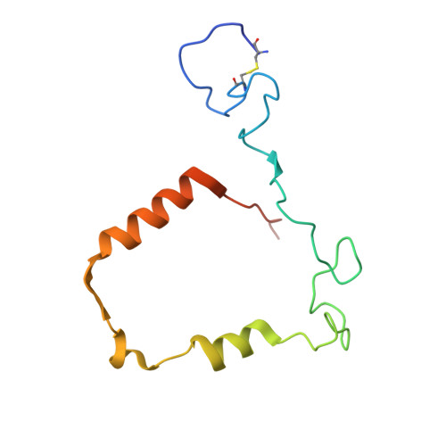
Halide binding by myeloperoxidase is regulated by access channel dynamics and charge interactions.

Leitgeb, U.Crha, R.Fegerl, I.Furtmuller, P.G.Oostenbrink, C.Pfanzagl, V.

(2025) Int J Biol Macromol : 148038-148038

  • DOI: https://doi.org/10.1016/j.ijbiomac.2025.148038
  • Primary Citation of Related Structures:  
    9QE3, 9QEX, 9QGA, 9QJ3, 9QJO, 9SDS

  • PubMed Abstract: 

    The heme enzyme myeloperoxidase is a key player in the innate immune defense. It uses hydrogen peroxide to produce bactericidal hypohalous acids from (pseudo)halides, foremost chloride, and thiocyanate in the neutrophil phagosome. However, the available structural data on the halide-binding site, the marked pH dependence of halide oxidation, and the atypical pK a of an active-site histidine 261 fail to fully account for the mechanism of halide oxidation by myeloperoxidase. In the present study, crystal structures of myeloperoxidase-halide complexes show that halides can integrate into the hydrogen-bonding network formed by conserved water molecules, without directly interacting with the deprotonated histidine at both acidic and neutral pH. Molecular dynamics simulations reveal that protonation of histidine 261 decreases active site rigidity and increases the flexibility of arginine 405. Together with the terminal residues of the myeloperoxidase heavy and light chains, arginine 405 contributes to halide transport into the active site. Kinetic analyses and simulations further demonstrate that sodium ions play a critical role as charge shields, enabling halides to traverse the negatively charged access channel, which represents a key bottleneck for halide binding. Thus, halide access to the active site is governed by a complex interplay of electrostatic interactions involving both solvent ions and charged amino acid residues.


  • Organizational Affiliation
    • Institute of Biochemistry, Department of Natural Sciences and Sustainable Resources, BOKU University, Vienna, Muthgasse 18, Vienna, Austria.

Macromolecules
Find similar proteins by:  (by identity cutoff)  |  3D Structure
Entity ID: 1
MoleculeChains Sequence LengthOrganismDetailsImage
Myeloperoxidase heavy chainA [auth B],
B [auth D]
467Homo sapiensMutation(s): 0 
EC: 1.11.2.2
UniProt & NIH Common Fund Data Resources
Find proteins for P05164 (Homo sapiens)
Explore P05164 
Go to UniProtKB:  P05164
PHAROS:  P05164
GTEx:  ENSG00000005381 
Entity Groups  
Sequence Clusters30% Identity50% Identity70% Identity90% Identity95% Identity100% Identity
UniProt GroupP05164
Glycosylation
Glycosylation Sites: 3Go to GlyGen: P05164-1
Sequence Annotations
Expand
  • Reference Sequence
Find similar proteins by:  (by identity cutoff)  |  3D Structure
Entity ID: 2
MoleculeChains Sequence LengthOrganismDetailsImage
Myeloperoxidase light chainC [auth A],
D [auth C]
114Homo sapiensMutation(s): 0 
EC: 1.11.2.2
UniProt & NIH Common Fund Data Resources
Find proteins for P05164 (Homo sapiens)
Explore P05164 
Go to UniProtKB:  P05164
PHAROS:  P05164
GTEx:  ENSG00000005381 
Entity Groups  
Sequence Clusters30% Identity50% Identity70% Identity90% Identity95% Identity100% Identity
UniProt GroupP05164
Sequence Annotations
Expand
  • Reference Sequence
Find similar proteins by:  (by identity cutoff)  |  3D Structure
Entity ID: 3
MoleculeChains Sequence LengthOrganismDetailsImage
Myeloperoxidase inhibitor SPIN
E, F
73Staphylococcus aureusMutation(s): 0 
Gene Names: SAOUHSC_00401
UniProt
Find proteins for Q2G0X2 (Staphylococcus aureus (strain NCTC 8325 / PS 47))
Explore Q2G0X2 
Go to UniProtKB:  Q2G0X2
Entity Groups  
Sequence Clusters30% Identity50% Identity70% Identity90% Identity95% Identity100% Identity
UniProt GroupQ2G0X2
Sequence Annotations
Expand
  • Reference Sequence
Oligosaccharides

Help

Entity ID: 4
MoleculeChains Length2D Diagram Glycosylation3D Interactions
2-acetamido-2-deoxy-beta-D-glucopyranose-(1-4)-2-acetamido-2-deoxy-beta-D-glucopyranose
G
2N-Glycosylation
Entity ID: 5
MoleculeChains Length2D Diagram Glycosylation3D Interactions
alpha-D-mannopyranose-(1-3)-[alpha-D-mannopyranose-(1-6)]beta-D-mannopyranose-(1-4)-2-acetamido-2-deoxy-beta-D-glucopyranose-(1-4)-[alpha-L-fucopyranose-(1-6)]2-acetamido-2-deoxy-beta-D-glucopyranose
H, I
6N-Glycosylation
Small Molecules
Ligands 4 Unique
IDChains Name / Formula / InChI Key2D Diagram3D Interactions
HEM (Subject of Investigation/LOI)
Query on HEM

Download Ideal Coordinates CCD File 
MA [auth B],
PA [auth D]
PROTOPORPHYRIN IX CONTAINING FE
C34 H32 Fe N4 O4
KABFMIBPWCXCRK-RGGAHWMASA-L
NAG
Query on NAG

Download Ideal Coordinates CCD File 
J [auth B],
NA [auth D],
OA [auth D]
2-acetamido-2-deoxy-beta-D-glucopyranose
C8 H15 N O6
OVRNDRQMDRJTHS-FMDGEEDCSA-N
BR
Query on BR

Download Ideal Coordinates CCD File 
AA [auth B]
AB [auth D]
BA [auth B]
BB [auth D]
CA [auth B]
AA [auth B],
AB [auth D],
BA [auth B],
BB [auth D],
CA [auth B],
CB [auth D],
DA [auth B],
DB [auth D],
EA [auth B],
EB [auth D],
FA [auth B],
GA [auth B],
GB [auth A],
HA [auth B],
HB [auth A],
IA [auth B],
IB [auth A],
JA [auth B],
JB [auth A],
K [auth B],
KA [auth B],
KB [auth A],
L [auth B],
LB [auth A],
M [auth B],
MB [auth A],
N [auth B],
NB [auth C],
O [auth B],
OB [auth C],
P [auth B],
PB [auth C],
Q [auth B],
QA [auth D],
QB [auth C],
R [auth B],
RA [auth D],
RB [auth C],
S [auth B],
SA [auth D],
SB [auth C],
T [auth B],
TA [auth D],
TB [auth F],
U [auth B],
UA [auth D],
V [auth B],
VA [auth D],
W [auth B],
WA [auth D],
X [auth B],
XA [auth D],
Y [auth B],
YA [auth D],
Z [auth B],
ZA [auth D]
BROMIDE ION
Br
CPELXLSAUQHCOX-UHFFFAOYSA-M
CA
Query on CA

Download Ideal Coordinates CCD File 
FB [auth D],
LA [auth B]
CALCIUM ION
Ca
BHPQYMZQTOCNFJ-UHFFFAOYSA-N
Modified Residues  1 Unique
IDChains TypeFormula2D DiagramParent
CSO
Query on CSO
A [auth B],
B [auth D]
L-PEPTIDE LINKINGC3 H7 N O3 SCYS
Experimental Data & Validation

Experimental Data

  • Method: X-RAY DIFFRACTION
  • Resolution: 2.18 Å
  • R-Value Free:  0.232 (Depositor), 0.232 (DCC) 
  • R-Value Work:  0.178 (Depositor), 0.182 (DCC) 
  • R-Value Observed: 0.181 (Depositor) 
Space Group: P 43 21 2
Unit Cell:
Length ( Å )Angle ( ˚ )
a = 110.878α = 90
b = 110.878β = 90
c = 240.703γ = 90
Software Package:
Software NamePurpose
REFMACrefinement
Aimlessdata scaling
XDSdata reduction
MOLREPphasing

Structure Validation

View Full Validation Report


Ligand Structure Quality Assessment 


Entry History & Funding Information

Deposition Data


Funding OrganizationLocationGrant Number
Austrian Science FundAustriaP33997
Austrian Science FundAustriaW1224

Revision History  (Full details and data files)

  • Version 1.0: 2025-10-15
    Type: Initial release