9OVY | pdb_00009ovy

Cryo-EM structure of human PRMT5:MEP50 in complex with SAH and compounds 16-19F and HJL-1


Experimental Data Snapshot

  • Method: ELECTRON MICROSCOPY
  • Resolution: 3.10 Å
  • Aggregation State: PARTICLE 
  • Reconstruction Method: SINGLE PARTICLE 

wwPDB Validation 3D Report Full Report

Validation slider image for 9OVY

This is version 2.0 of the entry. See complete history

Literature

Cryo-EM structure-based discovery of etravirine as a specific inhibitor of PRMT5/pICln protein-protein interaction for prostate cancer treatment.

Chi, Z.Xu, X.Shen, Z.Deng, X.Elzey, B.D.Li, C.Jiang, W.Hu, C.D.

(2026) J Enzyme Inhib Med Chem 41: 2727844-2727844

  • DOI: https://doi.org/10.1080/14756366.2026.2727844
  • Primary Citation Related Structures: 
    9OVY, 9Z49, 9Z4A

  • PubMed Abstract: 

    Protein arginine methyltransferase 5 (PRMT5) is overexpressed in many cancers and correlates with poor patient survival. In prostate cancer, PRMT5 cooperates with its cofactor pICln to promote tumour growth by epigenetically activating androgen receptor (AR) expression. Using a near-atomic cryo-EM structure of PRMT5/MEP50/pICln complex, we identified a previously undefined, pICln-specific protein-protein interaction (PPI) interface on PRMT5, termed P4I. Structure-based virtual screening identified the FDA-approved compound etravirine as a binder to this site. BiFC, Co-IP, and PLA assays confirmed that etravirine disrupts PRMT5/pICln interaction. A cryo-EM structure of PRMT5/MEP50/etravirine further validated on-target binding at P4I. Functionally, etravirine reduced prostate cancer cell proliferation, inhibited tumour growth, and downregulated AR and AR-V7 expression in cells and in mouse models. These results demonstrate that the unique P4I interface is a promising therapeutic target and that etravirine serves as a proof-of-concept lead compound for exploring the potential of P4I-targeted strategies in prostate cancer.


  • Organizational Affiliation
    • Department of Chemistry, Purdue University, West Lafayette, Indiana, USA.

Macromolecule Content 

  • Total Structure Weight: 406.03 kDa 
  • Atom Count: 27,074 
  • Modeled Residue Count: 3,364 
  • Deposited Residue Count: 3,574 
  • Unique protein chains: 2

Macromolecules

Find similar proteins by:|  3D Structure
Entity ID: 1
MoleculeChains  Sequence LengthOrganismDetailsImage
Protein arginine N-methyltransferase 5A,
C [auth G],
D [auth E],
F [auth C]
637Homo sapiensMutation(s): 0 
Gene Names: PRMT5HRMT1L5IBP72JBP1SKB1
EC: 2.1.1.320
UniProt & NIH Common Fund Data Resources
Find proteins for O14744 (Homo sapiens)
Explore O14744 
Go to UniProtKB:  O14744
PHAROS:  O14744
GTEx:  ENSG00000100462 
Entity Groups
Sequence Clusters30% Identity50% Identity70% Identity90% Identity95% Identity100% Identity
UniProt GroupO14744
Sequence Annotations
Expand
Reference Sequence
Find similar proteins by:|  3D Structure
Entity ID: 2
MoleculeChains  Sequence LengthOrganismDetailsImage
Methylosome protein 50B,
E [auth F],
G [auth D]
342Homo sapiensMutation(s): 0 
Gene Names: WDR77MEP50WD45HKMT1069Nbla10071
UniProt & NIH Common Fund Data Resources
Find proteins for Q9BQA1 (Homo sapiens)
Explore Q9BQA1 
Go to UniProtKB:  Q9BQA1
PHAROS:  Q9BQA1
GTEx:  ENSG00000116455 
Entity Groups
Sequence Clusters30% Identity50% Identity70% Identity90% Identity95% Identity100% Identity
UniProt GroupQ9BQA1
Sequence Annotations
Expand
Reference Sequence

Small Molecules

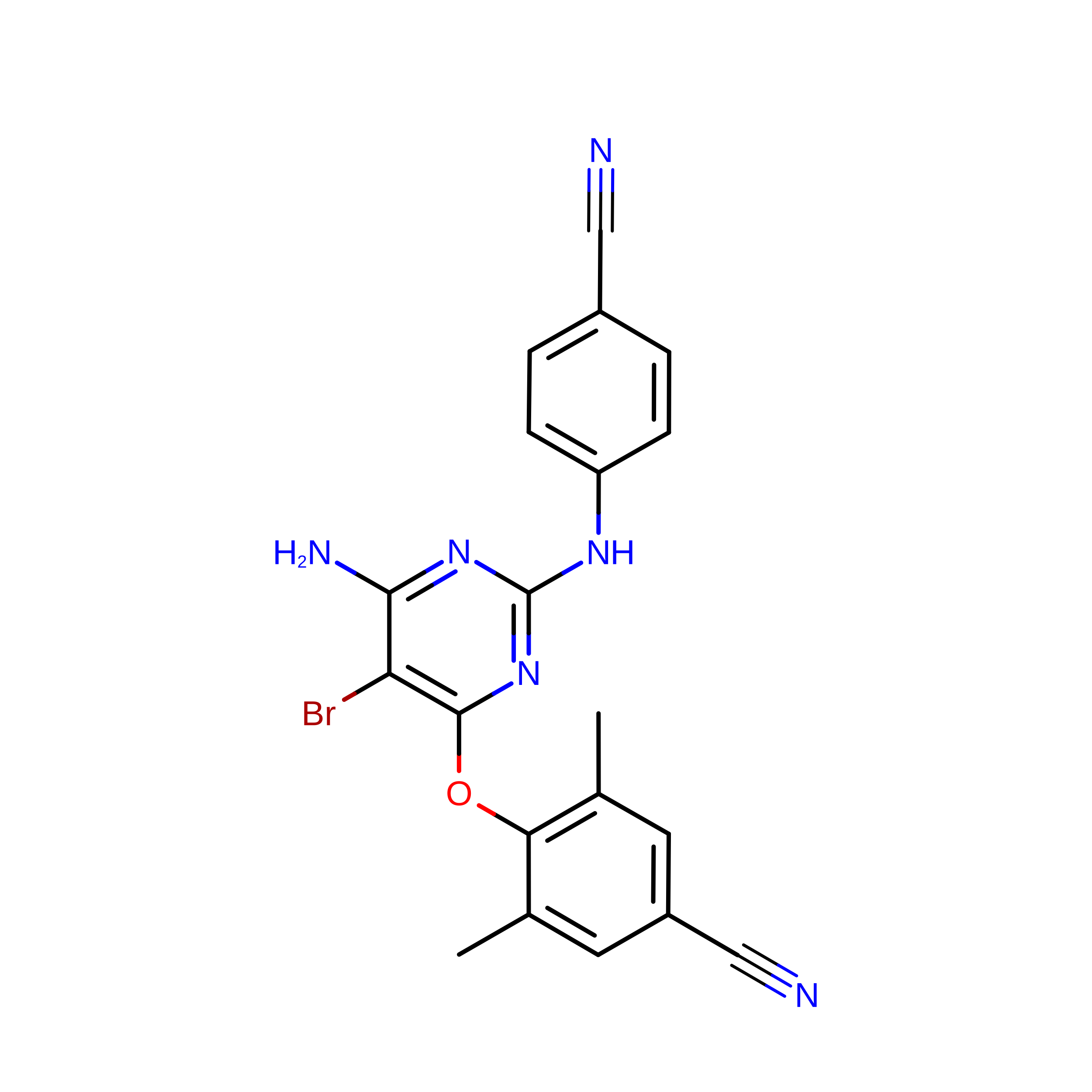
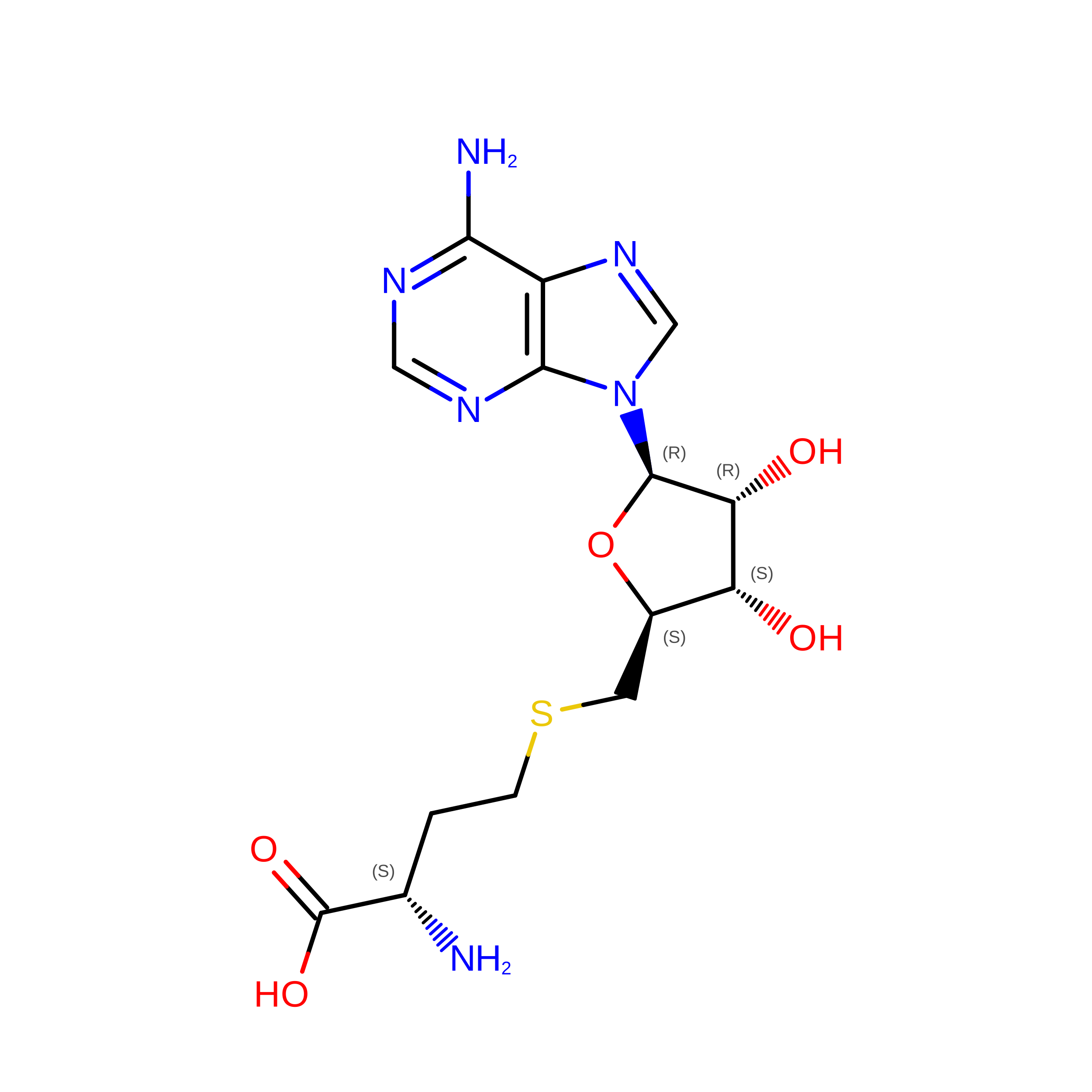
Ligands 3 Unique
IDChains Name / Formula / InChI Key2D Diagram3D Interactions
65B

Query on 65B


Download:Ideal Coordinates CCD File
H [auth A],
K [auth G],
N [auth E],
Q [auth C]
Etravirine
C20 H15 Br N6 O
PYGWGZALEOIKDF-UHFFFAOYSA-N
SAH

Query on SAH


Download:Ideal Coordinates CCD File
I [auth A],
L [auth G],
O [auth E],
R [auth C]
S-ADENOSYL-L-HOMOCYSTEINE
C14 H20 N6 O5 S
ZJUKTBDSGOFHSH-WFMPWKQPSA-N
A1CEU

Query on A1CEU


Download:Ideal Coordinates CCD File
J [auth A],
M [auth G],
P [auth E],
S [auth C]
8-chloro-1-[(2,4-diaminoquinazolin-7-yl)methyl]-3,4-dihydroquinolin-2(1H)-one
C18 H16 Cl N5 O
DVVSFRUZXCAZKM-UHFFFAOYSA-N

Experimental Data & Validation

Experimental Data

  • Method: ELECTRON MICROSCOPY
  • Resolution: 3.10 Å
  • Aggregation State: PARTICLE 
  • Reconstruction Method: SINGLE PARTICLE 
EM Software:
TaskSoftware PackageVersion
MODEL REFINEMENTPHENIX1.21.1_5286
RECONSTRUCTIONcryoSPARC

Structure Validation

View Full Validation Report


Entry History 

& Funding Information

Deposition Data


Funding OrganizationLocationGrant Number
National Institutes of Health/National Cancer Institute (NIH/NCI)United StatesR01CA212403

Revision History  (Full details and data files)

  • Version 1.0: 2026-06-10
    Type: Initial release
  • Version 2.0: 2026-09-16
    Changes: Data collection, Database references, Derived calculations, Non-polymer description, Structure summary