9MQW | pdb_00009mqw

Crystal structure of influenza virus N2 neuraminidase from A/Singapore/INFIMH-16-0019/2016 (H3N2)


Experimental Data Snapshot

  • Method: X-RAY DIFFRACTION
  • Resolution: 2.29 Å
  • R-Value Free: 
    0.214 (Depositor), 0.214 (DCC) 
  • R-Value Work: 
    0.175 (Depositor), 0.174 (DCC) 
  • R-Value Observed: 
    0.177 (Depositor) 

Starting Model: experimental
View more details

wwPDB Validation   3D Report Full Report


Ligand Structure Quality Assessment 


This is version 1.0 of the entry. See complete history


Literature

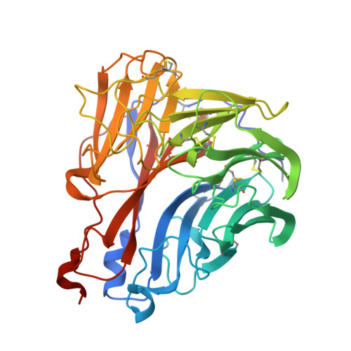
Structure and function of a cross-neutralizing influenza neuraminidase antibody that accommodates recent N2 NA Asn245 glycosylation.

Zhu, X.Khalil, A.M.Piepenbrink, M.S.Yu, W.Ma, Y.Martinez-Sobrido, L.Wilson, I.A.Kobie, J.J.

(2025) bioRxiv 

  • DOI: https://doi.org/10.1101/2025.06.30.662356
  • Primary Citation of Related Structures:  
    9MQV, 9MQW

  • PubMed Abstract: 

    Monoclonal antibodies (mAbs) that recognize and inhibit a diverse range of influenza viruses, although relatively rare, have been isolated following infection or vaccination. Study of their ontology and mechanisms of action informs universal vaccine and therapeutic development. We have previously described a potent and broad neuraminidase (NA)-neutralizing human mAb, 1122A11, that neutralizes a wide range of H3N2 viruses. Here, further characterization of 1122A11 reveals reactivity to cross-group influenza A virus NAs, including group-1 N1 and N8, and group-2 N2 and N3 NAs. Recent H3N2 viruses have acquired Asn245 glycosylation on the active site rim. Crystal structures of an N2 NA from A/Singapore/INFIMH-16-0019/2016 (H3N2) at 2.3 Å (apo) and 2.2 Å (Fab bound) resolution showed that 1122A11 binding causes local changes to the periphery of NA active site to accommodate the glycan. The CDRH3 of 1122A11 inserts into the active site and mimics the substrate sialic acid. We then determined that the ability of 1122A11 to protect from lethal challenge in mice is not dependent on Fc-effector function. These results highlight the therapeutic potential of 1122A11 as a broad protective anti-viral and reinforce pursuit of immunogen development of NA antibodies toward achieving more universal influenza protection.


  • Organizational Affiliation
    • Department of Integrative Structural and Computational Biology, The Scripps Research Institute, La Jolla, CA 92037, USA.

Macromolecules
Find similar proteins by:  (by identity cutoff)  |  3D Structure
Entity ID: 1
MoleculeChains Sequence LengthOrganismDetailsImage
Neuraminidase395Influenza A virusMutation(s): 0 
Gene Names: NA
EC: 3.2.1.18
UniProt
Find proteins for A0A193ACB1 (Influenza A virus)
Explore A0A193ACB1 
Go to UniProtKB:  A0A193ACB1
Entity Groups  
Sequence Clusters30% Identity50% Identity70% Identity90% Identity95% Identity100% Identity
UniProt GroupA0A193ACB1
Glycosylation
Glycosylation Sites: 6
Sequence Annotations
Expand
  • Reference Sequence
Oligosaccharides

Help

Entity ID: 2
MoleculeChains Length2D Diagram Glycosylation3D Interactions
2-acetamido-2-deoxy-beta-D-glucopyranose-(1-4)-2-acetamido-2-deoxy-beta-D-glucopyranose
B, C, D, E
2N-Glycosylation
Glycosylation Resources
GlyTouCan:  G42666HT
GlyCosmos:  G42666HT
GlyGen:  G42666HT
Entity ID: 3
MoleculeChains Length2D Diagram Glycosylation3D Interactions
alpha-D-mannopyranose-(1-2)-alpha-D-mannopyranose-(1-2)-alpha-D-mannopyranose-(1-3)-[alpha-D-mannopyranose-(1-3)-[alpha-D-mannopyranose-(1-6)]alpha-D-mannopyranose-(1-6)]beta-D-mannopyranose-(1-4)-2-acetamido-2-deoxy-beta-D-glucopyranose-(1-4)-2-acetamido-2-deoxy-beta-D-glucopyranose
F
9N-Glycosylation
Glycosylation Resources
GlyTouCan:  G68668TB
GlyCosmos:  G68668TB
GlyGen:  G68668TB
Small Molecules
Ligands 3 Unique
IDChains Name / Formula / InChI Key2D Diagram3D Interactions
NAG
Query on NAG

Download Ideal Coordinates CCD File 
I [auth A]2-acetamido-2-deoxy-beta-D-glucopyranose
C8 H15 N O6
OVRNDRQMDRJTHS-FMDGEEDCSA-N
SO4
Query on SO4

Download Ideal Coordinates CCD File 
H [auth A]
J [auth A]
K [auth A]
L [auth A]
M [auth A]
H [auth A],
J [auth A],
K [auth A],
L [auth A],
M [auth A],
N [auth A],
O [auth A],
P [auth A],
Q [auth A],
R [auth A],
S [auth A]
SULFATE ION
O4 S
QAOWNCQODCNURD-UHFFFAOYSA-L
CA
Query on CA

Download Ideal Coordinates CCD File 
G [auth A]CALCIUM ION
Ca
BHPQYMZQTOCNFJ-UHFFFAOYSA-N
Experimental Data & Validation

Experimental Data

  • Method: X-RAY DIFFRACTION
  • Resolution: 2.29 Å
  • R-Value Free:  0.214 (Depositor), 0.214 (DCC) 
  • R-Value Work:  0.175 (Depositor), 0.174 (DCC) 
  • R-Value Observed: 0.177 (Depositor) 
Space Group: I 4 2 2
Unit Cell:
Length ( Å )Angle ( ˚ )
a = 137.32α = 90
b = 137.32β = 90
c = 155.516γ = 90
Software Package:
Software NamePurpose
PHENIXrefinement
HKL-3000data reduction
HKL-3000data scaling
PHASERphasing

Structure Validation

View Full Validation Report


Ligand Structure Quality Assessment 


Entry History & Funding Information

Deposition Data


Funding OrganizationLocationGrant Number
National Institutes of Health/National Institute Of Allergy and Infectious Diseases (NIH/NIAID)United States75N93019C00051

Revision History  (Full details and data files)

  • Version 1.0: 2025-07-23
    Type: Initial release