


STRUCTURE of protein XAN60475 at 1.31 angstroms resolution
Yu, H.To be published.
Experimental Data Snapshot
Starting Model: in silico
View more details
Entity ID: 1 | |||||
|---|---|---|---|---|---|
| Molecule | Chains | Sequence Length | Organism | Details | Image |
| Thymidylate synthase | 238 | Phage #D | Mutation(s): 0 Gene Names: 308Ecol101PP_00194 | 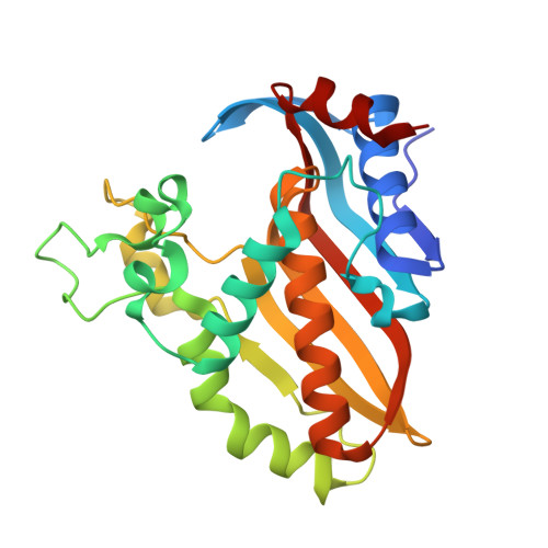 | |
UniProt | |||||
Find proteins for Q7Y590 (Escherichia phage RB69) Explore Q7Y590 Go to UniProtKB: Q7Y590 | |||||
Entity Groups | |||||
| Sequence Clusters | 30% Identity50% Identity70% Identity90% Identity95% Identity100% Identity | ||||
| UniProt Group | Q7Y590 | ||||
Sequence AnnotationsExpand | |||||
| |||||
| Ligands 3 Unique | |||||
|---|---|---|---|---|---|
| ID | Chains | Name / Formula / InChI Key | 2D Diagram | 3D Interactions | |
| FMN (Subject of Investigation/LOI) Query on FMN | B [auth A] | FLAVIN MONONUCLEOTIDE C17 H21 N4 O9 P FVTCRASFADXXNN-SCRDCRAPSA-N |  | ||
| DCM (Subject of Investigation/LOI) Query on DCM | C [auth A] | 2'-DEOXYCYTIDINE-5'-MONOPHOSPHATE C9 H14 N3 O7 P NCMVOABPESMRCP-SHYZEUOFSA-N |  | ||
| GOL (Subject of Investigation/LOI) Query on GOL | D [auth A] | GLYCEROL C3 H8 O3 PEDCQBHIVMGVHV-UHFFFAOYSA-N |  | ||
| Length ( Å ) | Angle ( ˚ ) |
|---|---|
| a = 64.6 | α = 90 |
| b = 64.6 | β = 90 |
| c = 230.21 | γ = 120 |
| Software Name | Purpose |
|---|---|
| PHENIX | refinement |
| XSCALE | data scaling |
| XDS | data reduction |
| PHENIX | phasing |
| Funding Organization | Location | Grant Number |
|---|---|---|
| Not funded | -- |


