Crystal engineering of the hepatoma-derived growth factor-related protein 2 PWWP domain towards crystallographic fragment screening.
Vantieghem, T., Osipov, E.M., Beelen, S., Strelkov, S.V.(2025) Acta Crystallogr F Struct Biol Commun 81: 358-364
- PubMed: 40709919
- DOI: https://doi.org/10.1107/S2053230X25006302
- Primary Citation of Related Structures:
9I7A, 9I7B, 9I7C, 9I7D, 9I7E, 9I7F, 9I7G, 9I7H, 9I7I, 9I7J - PubMed Abstract:
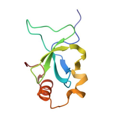
Hepatoma-derived growth factor-related protein 2 (HRP-2) is a member of the HDGF-related protein family, which has been linked to multiple malignancies. A defining feature of this protein family is the presence of an N-terminal PWWP domain, which enables binding to nucleosomes carrying a dimethylation or trimethylation marker on residue Lys36 of histone H3. To support the rational design of small-molecule drugs that bind to the PWWP domain, crystallographic fragment screening was chosen. A critical requirement for such screening is the ability to reliably produce large batches of high-quality crystals, ideally grown under low-salt conditions to prevent the precipitation of drug-like fragments during crystal soaking. Initial crystallization of the wild-type (WT) HRP-2 PWWP domain only produced crystals under high-salt conditions and these significantly lost diffraction quality over two weeks. It was hypothesized that these complications were caused by oxidation of the solvent-exposed Cys64 residue. To overcome these difficulties, a Cys64Ser mutant was produced. This mutation revealed a substantially improved crystallization propensity, as eight crystal forms could be obtained and resolved versus two forms for the WT. Moreover, the mutant crystals could be grown in PEG-based low ionic strength conditions which are optimal for fragment soaking. Finally, the crystals did not lose their diffraction quality for up to six months. Importantly, systematic analysis of all obtained X-ray structures revealed that the Cys64/Ser64 residue lies at a key lattice interface which is conserved across all crystal forms. This suggests that even minor chemical changes at this position could disrupt important intermolecular contacts, explaining the demonstrated major benefit of the introduced mutation. The presented data underpin the substitution of surface-exposed cysteines as a general strategy to enhance protein crystallization and diffraction quality. Ultimately, the results presented here were pivotal to the successful execution of the crystallographic fragment-screening campaign with the HRP-2 PWWP domain.
- Biocrystallography, Department of Pharmaceutical and Pharmacological Sciences, KU Leuven, Leuven, Belgium.
Organizational Affiliation: