


Structure of YIUA from Yersinia ruckeri with Iron and nitrilotriacetic acid
Thompson, S., Thomsen, E., Duhme-Klair, A., Butler, A., Grogan, G.To be published.
Experimental Data Snapshot
Starting Model: experimental
View more details
Entity ID: 1 | |||||
|---|---|---|---|---|---|
| Molecule | Chains | Sequence Length | Organism | Details | Image |
| Periplasmic substrate-binding transport protein | 347 | Yersinia ruckeri | Mutation(s): 0 Gene Names: CSF007_11785 | 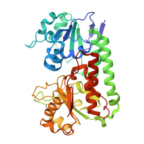 | |
UniProt | |||||
Find proteins for A0A085U4N5 (Yersinia ruckeri) Explore A0A085U4N5 Go to UniProtKB: A0A085U4N5 | |||||
Entity Groups | |||||
| Sequence Clusters | 30% Identity50% Identity70% Identity90% Identity95% Identity100% Identity | ||||
| UniProt Group | A0A085U4N5 | ||||
Sequence AnnotationsExpand | |||||
| |||||
| Ligands 2 Unique | |||||
|---|---|---|---|---|---|
| ID | Chains | Name / Formula / InChI Key | 2D Diagram | 3D Interactions | |
| NTA (Subject of Investigation/LOI) Query on NTA | D [auth A], F [auth B], H [auth C] | NITRILOTRIACETIC ACID C6 H9 N O6 MGFYIUFZLHCRTH-UHFFFAOYSA-N |  | ||
| FE (Subject of Investigation/LOI) Query on FE | E [auth A], G [auth B], I [auth C] | FE (III) ION Fe VTLYFUHAOXGGBS-UHFFFAOYSA-N |  | ||
| Length ( Å ) | Angle ( ˚ ) |
|---|---|
| a = 39.146 | α = 101.36 |
| b = 59.386 | β = 94.49 |
| c = 101.171 | γ = 100.54 |
| Software Name | Purpose |
|---|---|
| REFMAC | refinement |
| XDS | data reduction |
| SCALA | data scaling |
| MOLREP | phasing |
| Funding Organization | Location | Grant Number |
|---|---|---|
| Not funded | -- |


