9G53 | pdb_00009g53

The structure of Aspergillus fumigatus UDP-GlcNAc pyrophosphorylase in complex with a fragment


Experimental Data Snapshot

  • Method: X-RAY DIFFRACTION
  • Resolution: 1.96 Å
  • R-Value Free: 
    0.263 (Depositor), 0.264 (DCC) 
  • R-Value Work: 
    0.216 (Depositor), 0.217 (DCC) 
  • R-Value Observed: 
    0.218 (Depositor) 

Starting Model: experimental
View more details

wwPDB Validation   3D Report Full Report


Ligand Structure Quality Assessment 


This is version 1.0 of the entry. See complete history


Literature

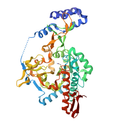
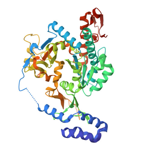
Exploration of starting points for the chemical validation of UDP-N-acetylglucosamine pyrophosphorylase in Aspergillus fumigatus

Yan, K.Stanley, M.Raimi, O.Kowalski, B.Gurvic, D.Grillenberger, S.Chen, X.Ferenbach, A.T.Dorfmueller, H.van Aalten, D.M.F.

To be published.

Macromolecules
Find similar proteins by:  (by identity cutoff)  |  3D Structure
Entity ID: 1
MoleculeChains Sequence LengthOrganismDetailsImage
UDP-N-acetylglucosamine diphosphorylaseA [auth B],
B [auth A]
484Aspergillus fumigatusMutation(s): 0 
Gene Names: KXV57_008857
EC: 2.7.7.23
UniProt
Find proteins for A0A229XUD0 (Aspergillus fumigatus)
Explore A0A229XUD0 
Go to UniProtKB:  A0A229XUD0
Entity Groups  
Sequence Clusters30% Identity50% Identity70% Identity90% Identity95% Identity100% Identity
UniProt GroupA0A229XUD0
Sequence Annotations
Expand
  • Reference Sequence
Find similar proteins by:  (by identity cutoff)  |  3D Structure
Entity ID: 2
MoleculeChains Sequence LengthOrganismDetailsImage
UDP-N-acetylglucosamine diphosphorylase
C, D
484Aspergillus fumigatusMutation(s): 0 
Gene Names: KXV57_008857
EC: 2.7.7.23
UniProt
Find proteins for A0A229XUD0 (Aspergillus fumigatus)
Explore A0A229XUD0 
Go to UniProtKB:  A0A229XUD0
Entity Groups  
Sequence Clusters30% Identity50% Identity70% Identity90% Identity95% Identity100% Identity
UniProt GroupA0A229XUD0
Sequence Annotations
Expand
  • Reference Sequence
Small Molecules
Ligands 4 Unique
IDChains Name / Formula / InChI Key2D Diagram3D Interactions
GN1
Query on GN1

Download Ideal Coordinates CCD File 
E [auth B],
I [auth A]
2-acetamido-2-deoxy-1-O-phosphono-alpha-D-glucopyranose
C8 H16 N O9 P
FZLJPEPAYPUMMR-FMDGEEDCSA-N
71G
Query on 71G

Download Ideal Coordinates CCD File 
G [auth B],
L [auth D],
M [auth D]
3-aminobenzonitrile
C7 H6 N2
NJXPYZHXZZCTNI-UHFFFAOYSA-N
CL
Query on CL

Download Ideal Coordinates CCD File 
K [auth C]CHLORIDE ION
Cl
VEXZGXHMUGYJMC-UHFFFAOYSA-M
MG
Query on MG

Download Ideal Coordinates CCD File 
H [auth B]MAGNESIUM ION
Mg
JLVVSXFLKOJNIY-UHFFFAOYSA-N
Modified Residues  1 Unique
IDChains TypeFormula2D DiagramParent
YCM
Query on YCM
A [auth B],
B [auth A]
L-PEPTIDE LINKINGC5 H10 N2 O3 SCYS
Experimental Data & Validation

Experimental Data

  • Method: X-RAY DIFFRACTION
  • Resolution: 1.96 Å
  • R-Value Free:  0.263 (Depositor), 0.264 (DCC) 
  • R-Value Work:  0.216 (Depositor), 0.217 (DCC) 
  • R-Value Observed: 0.218 (Depositor) 
Space Group: P 1 21 1
Unit Cell:
Length ( Å )Angle ( ˚ )
a = 55.67α = 90
b = 136.77β = 90.03
c = 142.55γ = 90
Software Package:
Software NamePurpose
PHENIXrefinement
XDSdata reduction
Aimlessdata scaling
PHASERphasing

Structure Validation

View Full Validation Report


Ligand Structure Quality Assessment 


Entry History & Funding Information

Deposition Data

  • Released Date: 2025-07-30 
  • Deposition Author(s): Yan, K.

Funding OrganizationLocationGrant Number
Medical Research Council (MRC, United Kingdom)United KingdomMR/V001094/1

Revision History  (Full details and data files)

  • Version 1.0: 2025-07-30
    Type: Initial release