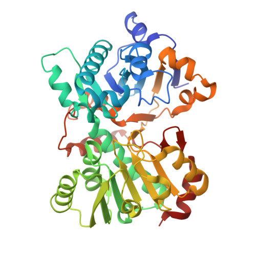
9E50 | pdb_00009e50

TAD from Carmabin Biosynthetic Pathway in complex with NAD - Crystal Form 2


Experimental Data Snapshot

  • Method: X-RAY DIFFRACTION
  • Resolution: 1.68 Å
  • R-Value Free: 
    0.230 (Depositor), 0.228 (DCC) 
  • R-Value Work: 
    0.201 (Depositor), 0.201 (DCC) 
  • R-Value Observed: 
    0.202 (Depositor) 

Starting Model: experimental
View more details

wwPDB Validation   3D Report Full Report


Ligand Structure Quality Assessment 


This is version 1.3 of the entry. See complete history


Literature

Designing small molecules targeting a cryptic RNA binding site through base displacement.

Olenginski, L.T.Wierzba, A.J.Laursen, S.P.Batey, R.T.

(2025) Nat Chem Biol 

  • DOI: https://doi.org/10.1038/s41589-025-02018-8
  • Primary Citation of Related Structures:  
    9E50, 9E5H, 9E5I, 9E5J, 9E5K, 9E5L, 9E5M, 9E5O, 9E5P, 9E5Q, 9E5R, 9E5S, 9E5T, 9ELR, 9MFH

  • PubMed Abstract: 

    Most RNA-binding small molecules have limited solubility, weak affinity and/or lack of specificity, restricting the medicinal chemistry often required for lead compound discovery. We reasoned that conjugation of these unfavorable ligands to a suitable 'host' molecule can solubilize the 'guest' and deliver it site-specifically to an RNA of interest to resolve these issues. Using this framework, we designed a small-molecule library that was hosted by cobalamin (Cbl) to interact with the Cbl riboswitch through a common base displacement mechanism. Combining in vitro binding, cell-based assays, chemoinformatic modeling and structure-based design, we unmasked a cryptic binding site within the riboswitch that was exploited to discover compounds that have affinity exceeding the native ligand, antagonize riboswitch function or bear no resemblance to Cbl. These data demonstrate how a privileged biphenyl-like scaffold effectively targets RNA by optimizing π-stacking interactions within the binding pocket.


  • Organizational Affiliation
    • Department of Biochemistry, University of Colorado Boulder, Boulder, CO, USA.

Macromolecules
Find similar proteins by:  (by identity cutoff)  |  3D Structure
Entity ID: 1
MoleculeChains Sequence LengthOrganismDetailsImage
Amino acid adenylation domain protein
A, B
399Moorena producens 3LMutation(s): 0 
Gene Names: LYNGBM3L_66040
UniProt
Find proteins for F4Y2B0 (Moorena producens 3L)
Explore F4Y2B0 
Go to UniProtKB:  F4Y2B0
Entity Groups  
Sequence Clusters30% Identity50% Identity70% Identity90% Identity95% Identity100% Identity
UniProt GroupF4Y2B0
Sequence Annotations
Expand
  • Reference Sequence
Experimental Data & Validation

Experimental Data

  • Method: X-RAY DIFFRACTION
  • Resolution: 1.68 Å
  • R-Value Free:  0.230 (Depositor), 0.228 (DCC) 
  • R-Value Work:  0.201 (Depositor), 0.201 (DCC) 
  • R-Value Observed: 0.202 (Depositor) 
Space Group: P 21 21 21
Unit Cell:
Length ( Å )Angle ( ˚ )
a = 55.433α = 90
b = 72.813β = 90
c = 209.207γ = 90
Software Package:
Software NamePurpose
PHENIXrefinement
XDSdata reduction
XDSdata scaling
PHASERphasing

Structure Validation

View Full Validation Report


Ligand Structure Quality Assessment 


Entry History & Funding Information

Deposition Data


Funding OrganizationLocationGrant Number
National Institutes of Health/National Institute of Diabetes and Digestive and Kidney Disease (NIH/NIDDK)United StatesR01DK042303
National Institutes of Health/National Cancer Institute (NIH/NCI)United StatesF31CA265082

Revision History  (Full details and data files)

  • Version 1.0: 2025-02-26
    Type: Initial release
  • Version 1.1: 2025-03-26
    Changes: Database references
  • Version 1.2: 2025-05-14
    Changes: Database references
  • Version 1.3: 2025-09-24
    Changes: Database references