9C3K | pdb_00009c3k

Crystal structure of GDP-bound KRAS G12D/M67R: Suppressing G12D oncogenicity via second-site M67R mutation


Experimental Data Snapshot

  • Method: X-RAY DIFFRACTION
  • Resolution: 1.70 Å
  • R-Value Free: 
    0.209 (Depositor), 0.209 (DCC) 
  • R-Value Work: 
    0.170 (Depositor), 0.172 (DCC) 
  • R-Value Observed: 
    0.184 (Depositor) 

Starting Model: experimental
View more details

wwPDB Validation   3D Report Full Report


Ligand Structure Quality Assessment 


This is version 1.0 of the entry. See complete history


Literature

Comprehensive structure-function analysis reveals gain- and loss-of-function mechanisms impacting oncogenic KRAS activity.

Kwon, J.J.Dilly, J.Liu, S.Kim, E.Bian, Y.Dharmaiah, S.Tran, T.H.Kapner, K.S.Ly, S.H.Yang, X.Rabara, D.Waybright, T.J.Giacomelli, A.O.Hong, A.L.Misek, S.Wang, B.Ravi, A.Doench, J.G.Beroukhim, R.Lemke, C.T.Haigis, K.M.Esposito, D.Root, D.E.Nissley, D.V.Stephen, A.G.McCormick, F.Simanshu, D.K.Hahn, W.C.Aguirre, A.J.

(2024) bioRxiv 

  • DOI: https://doi.org/10.1101/2024.10.22.618529
  • Primary Citation of Related Structures:  
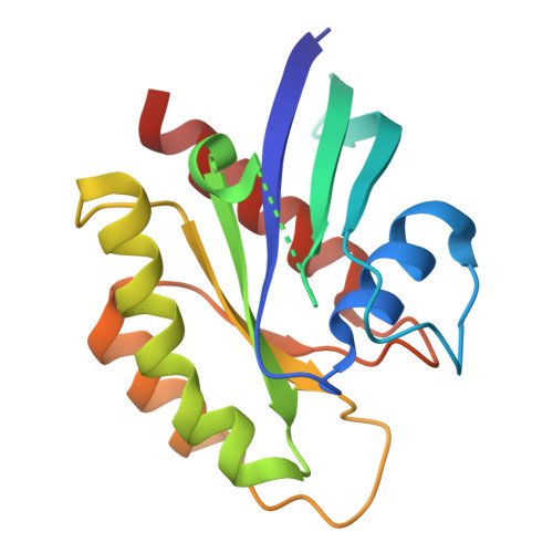
    9C3K, 9C3L, 9C3M, 9C3N, 9C3Q, 9C3R, 9C3V, 9C3Z, 9C40, 9C41, 9C43

  • PubMed Abstract: 

    To dissect variant-function relationships in the KRAS oncoprotein, we performed deep mutational scanning (DMS) screens for both wild-type and KRAS G12D mutant alleles. We defined the spectrum of oncogenic potential for nearly all possible KRAS variants, identifying several novel transforming alleles and elucidating a model to describe the frequency of KRAS mutations in human cancer as a function of transforming potential, mutational probability, and tissue-specific mutational signatures. Biochemical and structural analyses of variants identified in a KRAS G12D second-site suppressor DMS screen revealed that attenuation of oncogenic KRAS can be mediated by protein instability and conformational rigidity, resulting in reduced binding affinity to effector proteins, such as RAF and PI3-kinases, or reduced SOS-mediated nucleotide exchange activity. These studies define the landscape of single amino acid alterations that modulate the function of KRAS, providing a resource for the clinical interpretation of KRAS variants and elucidating mechanisms of oncogenic KRAS inactivation for therapeutic exploitation.


  • Organizational Affiliation
    • Department of Medical Oncology, Dana Farber Cancer Institute, Boston, MA, 02115, USA.

Macromolecules
Find similar proteins by:  (by identity cutoff)  |  3D Structure
Entity ID: 1
MoleculeChains Sequence LengthOrganismDetailsImage
Isoform 2B of GTPase KRas
A, B
170Homo sapiensMutation(s): 2 
Gene Names: KRASKRAS2RASK2
EC: 3.6.5.2
UniProt & NIH Common Fund Data Resources
Find proteins for P01116 (Homo sapiens)
Go to UniProtKB:  P01116
PHAROS:  P01116
GTEx:  ENSG00000133703 
Entity Groups  
Sequence Clusters30% Identity50% Identity70% Identity90% Identity95% Identity100% Identity
UniProt GroupP01116-2
Sequence Annotations
Expand
  • Reference Sequence
Experimental Data & Validation

Experimental Data

  • Method: X-RAY DIFFRACTION
  • Resolution: 1.70 Å
  • R-Value Free:  0.209 (Depositor), 0.209 (DCC) 
  • R-Value Work:  0.170 (Depositor), 0.172 (DCC) 
  • R-Value Observed: 0.184 (Depositor) 
Space Group: P 3
Unit Cell:
Length ( Å )Angle ( ˚ )
a = 84.63α = 90
b = 84.63β = 90
c = 41.76γ = 120
Software Package:
Software NamePurpose
XSCALEdata scaling
PHENIXrefinement
PDB_EXTRACTdata extraction
XDSdata reduction
PHENIXphasing

Structure Validation

View Full Validation Report


Ligand Structure Quality Assessment 


Entry History & Funding Information

Deposition Data


Funding OrganizationLocationGrant Number
National Institutes of Health/National Cancer Institute (NIH/NCI)United States75N91019D00024

Revision History  (Full details and data files)

  • Version 1.0: 2025-10-08
    Type: Initial release