8U2P | pdb_00008u2p

Crystal Structure of Glycine--tRNA ligase from Mycobacterium tuberculosis (G5A bound)


Experimental Data Snapshot

  • Method: X-RAY DIFFRACTION
  • Resolution: 2.25 Å
  • R-Value Free: 
    0.208 (Depositor), 0.210 (DCC) 
  • R-Value Work: 
    0.179 (Depositor), 0.180 (DCC) 
  • R-Value Observed: 
    0.180 (Depositor) 

Starting Model: experimental
View more details

wwPDB Validation   3D Report Full Report


Ligand Structure Quality Assessment 


This is version 1.2 of the entry. See complete history


Literature

Crystal structures of Mycobacterium tuberculosis and Mycobacterium thermoresistibile glycyl-tRNA synthetases in various liganded states.

Fenwick, M.K.DeRocher, A.E.Craig, J.K.Harmon, E.K.Seibold, S.Liu, L.Battaile, K.P.Barrett, L.K.Van Voorhis, W.C.Phan, I.Q.Staker, B.L.Subramanian, S.Lovell, S.Myler, P.J.

(2025) PLoS One 20: e0326500-e0326500

  • DOI: https://doi.org/10.1371/journal.pone.0326500
  • Primary Citation of Related Structures:  
    8SLD, 8SLF, 8SLG, 8T5N, 8U2P

  • PubMed Abstract: 

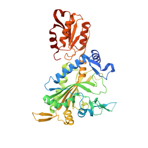
    Glycyl tRNA synthetases (GlyRSs) are prospective drug targets for combating Mycobacterium tuberculosis (Mtb) and cancer in humans. These synthetases are of the α2-subtype, with the ortholog in humans being dual targeted to the cytosol and mitochondria. Whereas the human enzyme has been structurally characterized previously in several liganded states, no structures of MtbGlyRS have thus far been reported. Here, we describe our recent work with MtbGlyRS and the closely-related Mycobacterium thermoresitibile GlyRS (MtrGlyRS), which progressed through all phases of the structural genomics pipeline, for the purpose of facilitating structure-based drug discovery. MtbGlyRS was expressed in Mycobacterium smegmatis and MtrGlyRS in Escherichia coli. Crystal structures are described for complexes of the two enzymes with adenosine monophosphate (AMP) and glycyl-sulfamoyl-adenylate (glycyl-AMS) at resolutions of 1.65/2.90 and 2.25/1.95 Å, respectively, and for MtrGlyRS in its apo state at 2.85 Å. Despite crystallizing in the dimeric state characteristic of many class II synthetases, the two enzymes elute predominantly as monomers during size exclusion chromatography. Strikingly, significant portions of the dimer interface and active site are unstructured in the MtrGlyRS apoenzyme crystal. AMP orders two tRNA recognition loops and a section of the insertion domain, and glycyl-AMS further stabilizes the structure, including the closure of a lid motif. Both the active and anticodon binding sites display structural differences with the human GlyRS and thus the collection of crystal structures should be useful for guiding drug development efforts targeting the various characterized structural states.


  • Organizational Affiliation

    Seattle Structural Genomics Center for Infectious Disease, Seattle, Washington, United States of America.


Macromolecules
Find similar proteins by:  (by identity cutoff)  |  3D Structure
Entity ID: 1
MoleculeChains Sequence LengthOrganismDetailsImage
Glycine--tRNA ligase476Mycobacterium tuberculosisMutation(s): 0 
Gene Names: glyQSglySRv2357cMTCY27.23MTCY98.26
EC: 6.1.1.14
UniProt
Find proteins for P9WFV7 (Mycobacterium tuberculosis (strain ATCC 25618 / H37Rv))
Explore P9WFV7 
Go to UniProtKB:  P9WFV7
Entity Groups  
Sequence Clusters30% Identity50% Identity70% Identity90% Identity95% Identity100% Identity
UniProt GroupP9WFV7
Sequence Annotations
Expand
  • Reference Sequence
Small Molecules
Ligands 6 Unique
IDChains Name / Formula / InChI Key2D Diagram3D Interactions
G5A (Subject of Investigation/LOI)
Query on G5A

Download Ideal Coordinates CCD File 
B [auth A]5'-O-(glycylsulfamoyl)adenosine
C12 H17 N7 O7 S
AMWPZASLDLLQFT-JJNLEZRASA-N
MES
Query on MES

Download Ideal Coordinates CCD File 
E [auth A]2-(N-MORPHOLINO)-ETHANESULFONIC ACID
C6 H13 N O4 S
SXGZJKUKBWWHRA-UHFFFAOYSA-N
IMD
Query on IMD

Download Ideal Coordinates CCD File 
C [auth A],
D [auth A]
IMIDAZOLE
C3 H5 N2
RAXXELZNTBOGNW-UHFFFAOYSA-O
EDO
Query on EDO

Download Ideal Coordinates CCD File 
F [auth A],
G [auth A],
H [auth A]
1,2-ETHANEDIOL
C2 H6 O2
LYCAIKOWRPUZTN-UHFFFAOYSA-N
CL
Query on CL

Download Ideal Coordinates CCD File 
J [auth A],
K [auth A]
CHLORIDE ION
Cl
VEXZGXHMUGYJMC-UHFFFAOYSA-M
MG
Query on MG

Download Ideal Coordinates CCD File 
I [auth A]MAGNESIUM ION
Mg
JLVVSXFLKOJNIY-UHFFFAOYSA-N
Experimental Data & Validation

Experimental Data

  • Method: X-RAY DIFFRACTION
  • Resolution: 2.25 Å
  • R-Value Free:  0.208 (Depositor), 0.210 (DCC) 
  • R-Value Work:  0.179 (Depositor), 0.180 (DCC) 
  • R-Value Observed: 0.180 (Depositor) 
Space Group: P 61 2 2
Unit Cell:
Length ( Å )Angle ( ˚ )
a = 124.293α = 90
b = 124.293β = 90
c = 131.383γ = 120
Software Package:
Software NamePurpose
PHENIXrefinement
Aimlessdata scaling
XDSdata reduction
PHASERphasing

Structure Validation

View Full Validation Report


Ligand Structure Quality Assessment 


Entry History & Funding Information

Deposition Data


Funding OrganizationLocationGrant Number
National Institutes of Health/National Institute Of Allergy and Infectious Diseases (NIH/NIAID)United States75N93022C00036
National Institutes of Health/Office of the DirectorUnited StatesS10OD030394

Revision History  (Full details and data files)

  • Version 1.0: 2023-09-13
    Type: Initial release
  • Version 1.1: 2024-04-10
    Changes: Source and taxonomy
  • Version 1.2: 2025-07-16
    Changes: Database references, Structure summary