4KGZ | pdb_00004kgz

The R state structure of E. coli ATCase with UTP and Magnesium bound


Experimental Data Snapshot

  • Method: X-RAY DIFFRACTION
  • Resolution: 2.40 Å
  • R-Value Free: 
    0.203 (Depositor), 0.207 (DCC) 
  • R-Value Work: 
    0.165 (Depositor), 0.170 (DCC) 
  • R-Value Observed: 
    0.167 (Depositor) 

Starting Model: experimental
View more details

wwPDB Validation 3D Report Full Report

Validation slider image for 4KGZ

Ligand Structure Quality Assessment 


This is version 1.2 of the entry. See complete history

Literature

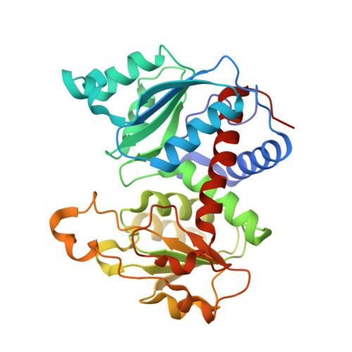
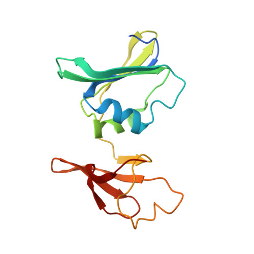
New Paradigm for Allosteric Regulation of Escherichia coli Aspartate Transcarbamoylase.

Cockrell, G.M.Zheng, Y.Guo, W.Peterson, A.W.Truong, J.K.Kantrowitz, E.R.

(2013) Biochemistry 52: 8036-8047

  • DOI: https://doi.org/10.1021/bi401205n
  • Primary Citation Related Structures: 
    4KGV, 4KGX, 4KGZ, 4KH0, 4KH1

  • PubMed Abstract: 

    For nearly 60 years, the ATP activation and the CTP inhibition of Escherichia coli aspartate transcarbamoylase (ATCase) has been the textbook example of allosteric regulation. We present kinetic data and five X-ray structures determined in the absence and presence of a Mg(2+) concentration within the physiological range. In the presence of 2 mM divalent cations (Mg(2+), Ca(2+), Zn(2+)), CTP does not significantly inhibit the enzyme, while the allosteric activation by ATP is enhanced. The data suggest that the actual allosteric inhibitor of ATCase in vivo is the combination of CTP, UTP, and a divalent cation, and the actual allosteric activator is a divalent cation with ATP or ATP and GTP. The structural data reveals that two NTPs can bind to each allosteric site with a divalent cation acting as a bridge between the triphosphates. Thus, the regulation of ATCase is far more complex than previously believed and calls many previous studies into question. The X-ray structures reveal that the catalytic chains undergo essentially no alternations; however, several regions of the regulatory chains undergo significant structural changes. Most significant is that the N-terminal region of the regulatory chains exists in different conformations in the allosterically activated and inhibited forms of the enzyme. Here, a new model of allosteric regulation is proposed.


  • Organizational Affiliation
    • Department of Chemistry, Boston College , Merkert Chemistry Center, 2609 Beacon Street, Chestnut Hill, MA 02467 U.S.A.

Macromolecule Content 

  • Total Structure Weight: 105.59 kDa 
  • Atom Count: 7,565 
  • Modeled Residue Count: 907 
  • Deposited Residue Count: 926 
  • Unique protein chains: 2

Macromolecules

Find similar proteins by:|  3D Structure
Entity ID: 1
MoleculeChains  Sequence LengthOrganismDetailsImage
Aspartate carbamoyltransferase
A, C
310Escherichia coli KO11FLMutation(s): 0 
Gene Names: pyrBEKO11_4066KO11_22860
EC: 2.1.3.2
Find similar proteins by:|  3D Structure
Entity ID: 2
MoleculeChains  Sequence LengthOrganismDetailsImage
Aspartate carbamoyltransferase regulatory chain
B, D
153Escherichia coli KO11FLMutation(s): 0 
Gene Names: pyrIEKO11_4067KO11_22855
EC: 2.1.3.2

Small Molecules

Ligands 4 Unique
IDChains Name / Formula / InChI Key2D Diagram3D Interactions
UTP

Query on UTP


Download:Ideal Coordinates CCD File
G [auth B],
H [auth B],
L [auth D],
M [auth D]
URIDINE 5'-TRIPHOSPHATE
C9 H15 N2 O15 P3
PGAVKCOVUIYSFO-XVFCMESISA-N
PAL

Query on PAL


Download:Ideal Coordinates CCD File
E [auth A],
J [auth C]
N-(PHOSPHONACETYL)-L-ASPARTIC ACID
C6 H10 N O8 P
ZZKNRXZVGOYGJT-VKHMYHEASA-N
ZN

Query on ZN


Download:Ideal Coordinates CCD File
F [auth B],
K [auth D]
ZINC ION
Zn
PTFCDOFLOPIGGS-UHFFFAOYSA-N
MG

Query on MG


Download:Ideal Coordinates CCD File
I [auth B],
N [auth D]
MAGNESIUM ION
Mg
JLVVSXFLKOJNIY-UHFFFAOYSA-N

Experimental Data & Validation

Experimental Data

  • Method: X-RAY DIFFRACTION
  • Resolution: 2.40 Å
  • R-Value Free:  0.203 (Depositor), 0.207 (DCC) 
  • R-Value Work:  0.165 (Depositor), 0.170 (DCC) 
  • R-Value Observed: 0.167 (Depositor) 
Space Group: P 3 2 1
Unit Cell:
Length ( Å )Angle ( ˚ )
a = 121.378α = 90
b = 121.378β = 90
c = 155.135γ = 120
Software Package:
Software NamePurpose
DENZOdata reduction
SCALEPACKdata scaling
PHENIXrefinement
PDB_EXTRACTdata extraction
HKL-2000data collection
HKL-2000data reduction
HKL-2000data scaling
PHENIXphasing

Structure Validation

View Full Validation Report


Ligand Structure Quality Assessment 


Entry History 

Deposition Data

Revision History  (Full details and data files)

  • Version 1.0: 2013-11-27
    Type: Initial release
  • Version 1.1: 2017-11-15
    Changes: Refinement description
  • Version 1.2: 2023-09-20
    Changes: Data collection, Database references, Derived calculations, Refinement description