Download Mendeley
Cryo-EM Structure of Siphophage Avs-1 Reveals a Conserved Virion Architecture with a Specialized Tripod-like Tail Tip for Host Recognition.
Liu, J., Li, C., Yang, X., Yang, F., Liu, H.(2026) Viruses 18
- PubMed: 42797842 Search on PubMedSearch on PubMed Central
- DOI: https://doi.org/10.3390/v18091011
- Primary Citation Related Structures:
43JF, 43JG, 43JH - PubMed Abstract:
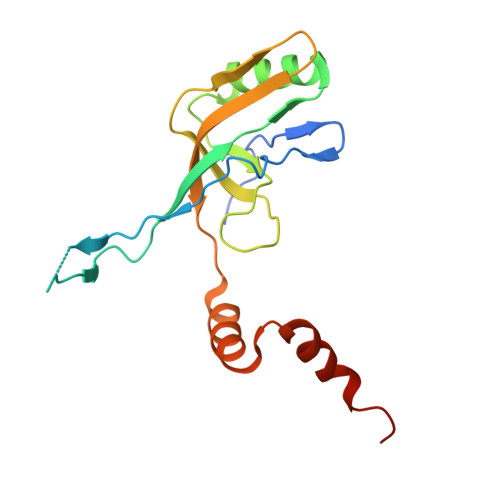
Bacteriophages are increasingly considered viable alternatives to antibiotics for controlling aquaculture pathogens, yet the structural basis of siphophage infection in aquatic bacteria remains largely unexplored. Here, we present the near-atomic-resolution cryo-electron microscopy structure of Avs-1, a siphophage infecting the freshwater aquaculture pathogen Aeromonas veronii . The capsid reconstruction at 2.98 Å and spike structure at 3.58 Å reveal a virion architecture broadly conserved among lambda-like siphophages, including an HK97-fold capsid, a canonical portal-neck complex, and a tail tube composed of approximately 26 stacked hexameric rings. However, the distal tail tip exhibits pronounced specialization, assembling into a C3-symmetric tripod-like core formed by the distal tail protein, hub protein, insertion protein, and the C-terminus of the tape measure protein. The trimeric spike protein gp43 extends from this platform and adopts a β-helical scaffold with a structurally divergent distal domain (D4), highlighting the structural diversity of phage tail-tip architectures. Together, these results reveal how a largely conserved virion framework is combined with a diversified distal tail apparatus, expanding the structural repertoire of characterized siphophages and providing a framework for investigating the molecular basis of phage-host interactions.
- Key Laboratory for Matter Microstructure and Function of Hunan Province, School of Physics and Electronics, Hunan Normal University, Changsha 410081, China.
Organizational Affiliation: