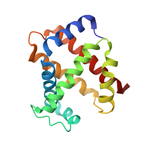
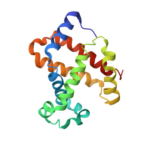
3D17 | pdb_00003d17

A triply ligated crystal structure of relaxed state human hemoglobin


Experimental Data Snapshot

  • Method: X-RAY DIFFRACTION
  • Resolution: 2.80 Å
  • R-Value Free: 
    0.297 (Depositor), 0.300 (DCC) 
  • R-Value Work: 
    0.232 (Depositor), 0.230 (DCC) 
  • R-Value Observed: 
    0.255 (Depositor) 

Starting Model: experimental
View more details

wwPDB Validation   3D Report Full Report


Ligand Structure Quality Assessment 


This is version 1.3 of the entry. See complete history


Literature

Structure of relaxed-state human hemoglobin: insight into ligand uptake, transport and release.

Jenkins, J.D.Musayev, F.N.Danso-Danquah, R.Abraham, D.J.Safo, M.K.

(2009) Acta Crystallogr D Biol Crystallogr 65: 41-48

  • DOI: https://doi.org/10.1107/S0907444908037256
  • Primary Citation of Related Structures:  
    3D17

  • PubMed Abstract: 

    Hemoglobin was one of the first protein structures to be determined by X-ray crystallography and served as a basis for the two-state MWC model for the mechanism of allosteric proteins. Since then, there has been an ongoing debate about whether Hb allostery involves the unliganded tense T state and the liganded relaxed R state or whether it involves the T state and an ensemble of liganded relaxed states. In fact, the former model is inconsistent with many functional observations, as well as the recent discoveries of several relaxed-state Hb structures such as RR2, R3 and R2. One school of thought has suggested the R2 state to be the physiologically relevant relaxed end state, with the R state mediating the T-->R2 transition. X-ray studies have been performed on human carbonmonoxy Hb at a resolution of 2.8 A. The ensuing liganded quaternary structure is different from previously reported liganded Hb structures. The distal beta-heme pocket is the largest when compared with other liganded Hb structures, partly owing to rotation of betaHis63(E7) out of the distal pocket, creating a ligand channel to the solvent. The structure also shows unusually smaller alpha- and beta-clefts. Results from this study taken in conjunction with previous findings suggest that multiple liganded Hb states with different quaternary structures may be involved in ligand uptake, stabilization, transport and release.


  • Organizational Affiliation
    • Department of Medicinal Chemistry, School of Pharmacy and Institute for Structural Biology and Drug Discovery, Virginia Commonwealth University, Richmond, VA 23219, USA.

Macromolecules
Find similar proteins by:  (by identity cutoff)  |  3D Structure
Entity ID: 1
MoleculeChains Sequence LengthOrganismDetailsImage
Hemoglobin subunit alpha
A, C
141Homo sapiensMutation(s): 0 
UniProt & NIH Common Fund Data Resources
Find proteins for P69905 (Homo sapiens)
Explore P69905 
Go to UniProtKB:  P69905
PHAROS:  P69905
Entity Groups  
Sequence Clusters30% Identity50% Identity70% Identity90% Identity95% Identity100% Identity
UniProt GroupP69905
Sequence Annotations
Expand
  • Reference Sequence
Find similar proteins by:  (by identity cutoff)  |  3D Structure
Entity ID: 2
MoleculeChains Sequence LengthOrganismDetailsImage
Hemoglobin subunit beta
B, D
146Homo sapiensMutation(s): 0 
UniProt & NIH Common Fund Data Resources
Find proteins for P68871 (Homo sapiens)
Explore P68871 
Go to UniProtKB:  P68871
PHAROS:  P68871
GTEx:  ENSG00000244734 
Entity Groups  
Sequence Clusters30% Identity50% Identity70% Identity90% Identity95% Identity100% Identity
UniProt GroupP68871
Sequence Annotations
Expand
  • Reference Sequence
Small Molecules
Ligands 4 Unique
IDChains Name / Formula / InChI Key2D Diagram3D Interactions
HEM
Query on HEM

Download Ideal Coordinates CCD File 
E [auth A],
J [auth B],
L [auth C],
N [auth D]
PROTOPORPHYRIN IX CONTAINING FE
C34 H32 Fe N4 O4
KABFMIBPWCXCRK-RGGAHWMASA-L
PO4
Query on PO4

Download Ideal Coordinates CCD File 
H [auth B],
I [auth B],
M [auth D]
PHOSPHATE ION
O4 P
NBIIXXVUZAFLBC-UHFFFAOYSA-K
MBN
Query on MBN

Download Ideal Coordinates CCD File 
F [auth A]TOLUENE
C7 H8
YXFVVABEGXRONW-UHFFFAOYSA-N
CMO
Query on CMO

Download Ideal Coordinates CCD File 
G [auth A],
K [auth B],
O [auth D]
CARBON MONOXIDE
C O
UGFAIRIUMAVXCW-UHFFFAOYSA-N
Experimental Data & Validation

Experimental Data

  • Method: X-RAY DIFFRACTION
  • Resolution: 2.80 Å
  • R-Value Free:  0.297 (Depositor), 0.300 (DCC) 
  • R-Value Work:  0.232 (Depositor), 0.230 (DCC) 
  • R-Value Observed: 0.255 (Depositor) 
Space Group: P 41 21 2
Unit Cell:
Length ( Å )Angle ( ˚ )
a = 87.282α = 90
b = 87.282β = 90
c = 174.466γ = 90
Software Package:
Software NamePurpose
d*TREKdata scaling
AMoREphasing
CNSrefinement
PDB_EXTRACTdata extraction
d*TREKdata reduction

Structure Validation

View Full Validation Report


Ligand Structure Quality Assessment 


Entry History 

Deposition Data

Revision History  (Full details and data files)

  • Version 1.0: 2008-06-03
    Type: Initial release
  • Version 1.1: 2011-07-13
    Changes: Version format compliance
  • Version 1.2: 2023-08-30
    Changes: Data collection, Database references, Derived calculations, Refinement description
  • Version 1.3: 2025-11-12
    Changes: Database references, Structure summary