32DH | pdb_000032dh

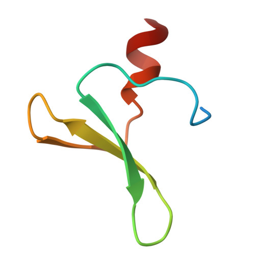
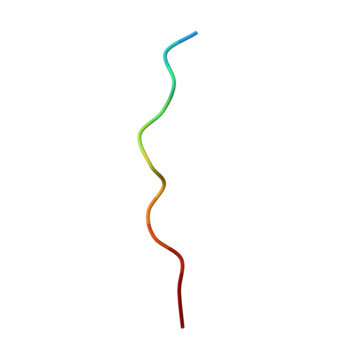
Crystal structure of YAP1(165-209) in complex with LATS2(511-522_C513S)


Experimental Data Snapshot

  • Method: X-RAY DIFFRACTION
  • Resolution: 1.03 Å
  • R-Value Free: 
    0.192 (Depositor), 0.192 (DCC) 
  • R-Value Work: 
    0.186 (Depositor), 0.187 (DCC) 
  • R-Value Observed: 
    0.187 (Depositor) 

Starting Model: experimental
View more details

wwPDB Validation 3D Report Full Report

Validation slider image for 32DH

This is version 1.0 of the entry. See complete history. 

Literature

Comparing YAP WW1 and TAZ WW : Similar Binding Sites but Different Stability and Conformational Dynamics.

Merlen, C., Mesrouze, Y., Chau, S., Diehl, B.A., Hinniger, A., Zimmermann, C., Meyerhofer, M., Fontana, P., Groebke, S., Hinrichs, J., Abdul Rahman, W., Gutmann, S., Fernandez, C., Petrovic, D., Chene, P.

(2026) Biomolecules 16

  • DOI: https://doi.org/10.3390/biom16091355
  • Primary Citation Related Structures: 
    32DH

  • PubMed Abstract: 

    The two paralogs YAP and TAZ act through TEAD transcription factors and bind PPxY motif proteins in the Hippo pathway via WW domains. YAP has up to two WW domains, whereas TAZ has one. Because YAP WW1 and TAZ WW are the most similar in sequence, they can be considered as corresponding modules in these two proteins. This study shows that, despite their similarity, they differ strongly in conformational stability and in how they bind flexible ligands. Nano-differential scanning fluorimetry, circular dichroism, and NMR indicate that both isolated domains populate partially folded or exchanging states in solution, but TAZ WW is more thermally stable and has a larger folded population. Peptide binding stabilizes both domains, producing sharper NMR signals. Surface plasmon resonance measurements with PPxY peptides show micromolar affinities and generally modest differences between YAP WW1 and TAZ WW , although these differences increase for conformationally plastic ligands. A high resolution LATS2:YAP WW1 crystal structure and molecular dynamics simulations suggest that preorganized peptides bind more tightly and less selectively, while flexible peptides incur larger binding penalties that dynamic YAP WW1 compensates more effectively. These observations indicate that differences in WW-domain stability contribute to subtle YAP/TAZ binding preferences despite conserved binding surfaces and evolutionary relatedness between the paralogs.


  • Organizational Affiliation: 
    • Oncology Disease Area, Novartis Biomedical Research, CH-4056 Basel, Switzerland.

Macromolecule Content 

  • Total Structure Weight: 7 kDa 
  • Atom Count: 524 
  • Modeled Residue Count: 55 
  • Deposited Residue Count: 62 
  • Unique protein chains: 2

Macromolecules

Find similar proteins by:|  3D Structure
Entity ID: 1
MoleculeChains  Sequence LengthOrganismDetailsImage
Transcriptional coactivator YAP149Homo sapiensMutation(s): 0 
Gene Names: YAP1, YAP65
UniProt & NIH Common Fund Data Resources
Find proteins for P46937 (Homo sapiens)
Explore P46937 
Go to UniProtKB:  P46937
PHAROS:  P46937
GTEx:  ENSG00000137693 
Entity Groups
Sequence Clusters30% Identity50% Identity70% Identity90% Identity95% Identity100% Identity
UniProt GroupP46937
Sequence Annotations
Expand
Reference Sequence
Find similar proteins by:  Sequence   |   3D Structure  
Entity ID: 2
MoleculeChains  Sequence LengthOrganismDetailsImage
Serine/threonine-protein kinase LATS2B [auth L]13Homo sapiensMutation(s): 2 
EC: 2.7.11.1
UniProt & NIH Common Fund Data Resources
Find proteins for Q9NRM7 (Homo sapiens)
Explore Q9NRM7 
Go to UniProtKB:  Q9NRM7
PHAROS:  Q9NRM7
GTEx:  ENSG00000150457 
Entity Groups
Sequence Clusters30% Identity50% Identity70% Identity90% Identity95% Identity100% Identity
UniProt GroupQ9NRM7
Sequence Annotations
Expand
Reference Sequence

Experimental Data & Validation

Experimental Data

  • Method: X-RAY DIFFRACTION
  • Resolution: 1.03 Å
  • R-Value Free:  0.192 (Depositor), 0.192 (DCC) 
  • R-Value Work:  0.186 (Depositor), 0.187 (DCC) 
  • R-Value Observed: 0.187 (Depositor) 
Space Group: P 31 2 1
Unit Cell:
Length ( Å )Angle ( ˚ )
a = 53.036α = 90
b = 53.036β = 90
c = 37.593γ = 120
Software Package:
Software NamePurpose
PHENIXrefinement
XDSdata reduction
Aimlessdata scaling
PHASERphasing

Structure Validation

View Full Validation Report


Entry History 

& Funding Information

Deposition Data


Funding OrganizationLocationGrant Number
Not funded--

Revision History  (Full details and data files)

  • Version 1.0: 2026-10-07
    Type: Initial release