 Download Mendeley
Molecular basis of PADI6 species-specific oligomerization in maternal regulatory complexes
Li, J., Deng, D.To be published.
Experimental Data Snapshot
wwPDB Validation 3D Report Full Report

Entity ID: 1 | |||||
|---|---|---|---|---|---|
| Molecule | Chains | Sequence Length | Organism | Details | Image |
| Inactive protein-arginine deiminase type-6 | 682 | Mus musculus | Mutation(s): 0 Gene Names: Padi6, Pad6, Padi5 | 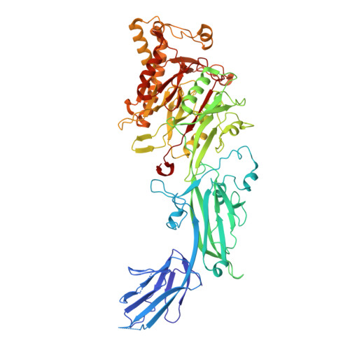 | |
UniProt & NIH Common Fund Data Resources | |||||
IMPC: MGI:2655198 | |||||
Entity Groups | |||||
| Sequence Clusters | 30% Identity50% Identity70% Identity90% Identity95% Identity100% Identity | ||||
| UniProt Group | Q8K3V4 | ||||
Sequence AnnotationsExpand | |||||
Reference Sequence | |||||
| Task | Software Package | Version |
|---|---|---|
| RECONSTRUCTION | cryoSPARC |
| Funding Organization | Location | Grant Number |
|---|---|---|
| National Natural Science Foundation of China (NSFC) | China | 31871449 |
