25IN | pdb_000025in

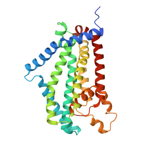
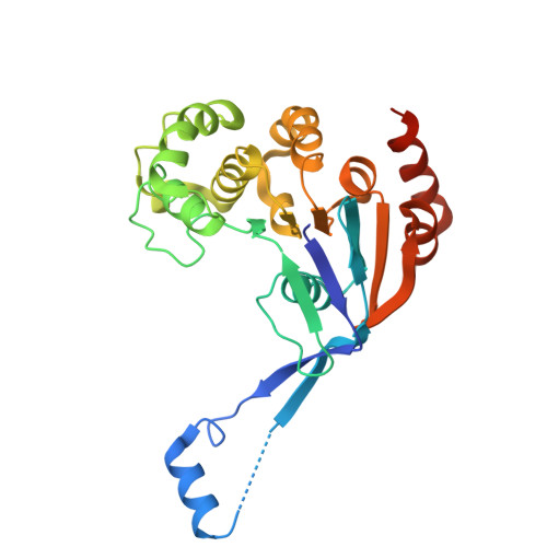
Mtb galactan transporter Rv3781/3783, open state


Experimental Data Snapshot

  • Method: ELECTRON MICROSCOPY
  • Resolution: 2.91 Å
  • Aggregation State: PARTICLE 
  • Reconstruction Method: SINGLE PARTICLE 

wwPDB Validation 3D Report Full Report

Validation slider image for 25IN

This is version 1.1 of the entry. See complete history. 

Literature

Recognition and mechanism of M. tuberculosis cell wall galactan transport.

Wei, C., Abrahams, K.A., Krisilia, V., Li, J., Gurcha, S.S., Derr, E., Hanisch, M., Kalscheuer, R., Besra, G.S., Rao, Z., Zhang, L.

(2026) Nat Commun 17

  • DOI: https://doi.org/10.1038/s41467-026-78496-8
  • Primary Citation Related Structures: 
    25IN, 25IO, 25IP

  • PubMed Abstract: 

    Mycobacterial-derived lipid-linked galactan (LLG) is an essential component of the Mycobacterial tuberculosis (Mtb) cell wall core arabinogalactan (AG), a well-established virulence factor and target of anti-tubercular drugs. Cross-membrane translocation of LLG is energetically unfavorable and is therefore catalyzed by WzmWzt, a prominent member of the polysaccharide ABC transporter sub-family. Here we present cryo-EM structures of WzmWzt Mtb to uncover critical steps of Mtb LLG translocation. Notably, densities suggestive of the mycobacterial-derived LLG substrates were observed in the structure of WzmWzt Mtb , indicating that the substrate may bind through two Wzm and two Wzt subunits. This proposed LLG binding is fulfilled by a fully upward extension of the Wzt gate helix, which serves as a critical step to prime Wzt for dimerization. The ATP-bound structure revealed a remarkable downward movement of the gate helix, together with the Wzt dimerization. The third, DDM/ATP-bound structure captured WztWzm Mtb in an intermediate state with two DDM molecules in the Wzm central cavity. Using two conditional mycobacterial mutants for functional analyses, we have identified residues on the Wzm N-terminal interface helix (IF N ) and on the Wzt helical domain are essential for LLG recognition and translocation in vivo. These data provide a clear structural basis for the development of Mtb cell wall targeted therapeutics for global tuberculosis treatment.


  • Organizational Affiliation: 
    • Shanghai Institute for Advanced Immunochemical Studies, School of Life Science and Technology, ShanghaiTech University, Shanghai, China.

Macromolecule Content 

  • Total Structure Weight: 128.71 kDa 
  • Atom Count: 8,584 
  • Modeled Residue Count: 1,044 
  • Deposited Residue Count: 1,106 
  • Unique protein chains: 2

Macromolecules

Find similar proteins by:|  3D Structure
Entity ID: 1
MoleculeChains  Sequence LengthOrganismDetailsImage
Probable O-antigen/lipopolysaccharide transport integral membrane protein ABC transporter RfbD
A, B
280Mycobacterium tuberculosis H37RvMutation(s): 0 
Gene Names: rfbD, Rv3783
UniProt
Find proteins for P72049 (Mycobacterium tuberculosis (strain ATCC 25618 / H37Rv))
Explore P72049 
Go to UniProtKB:  P72049
Entity Groups
Sequence Clusters30% Identity50% Identity70% Identity90% Identity95% Identity100% Identity
UniProt GroupP72049
Sequence Annotations
Expand
Reference Sequence
Find similar proteins by:|  3D Structure
Entity ID: 2
MoleculeChains  Sequence LengthOrganismDetailsImage
Probable O-antigen/lipopolysaccharide transport ATP-binding protein ABC transporter RfbE
C, D
273Mycobacterium tuberculosis H37RvMutation(s): 1 
Gene Names: rfbE, Rv3781
UniProt
Find proteins for P72047 (Mycobacterium tuberculosis (strain ATCC 25618 / H37Rv))
Explore P72047 
Go to UniProtKB:  P72047
Entity Groups
Sequence Clusters30% Identity50% Identity70% Identity90% Identity95% Identity100% Identity
UniProt GroupP72047
Sequence Annotations
Expand
Reference Sequence

Oligosaccharides

Help  
Entity ID: 3
MoleculeChains Length2D Diagram GlycosylationD Interactions
alpha-L-rhamnopyranose-(1-3)-2-acetamido-2-deoxy-1-O-phosphono-alpha-D-glucopyranose
E, F
2N/AN/A
Entity ID: 4
MoleculeChains Length2D Diagram GlycosylationD Interactions
beta-D-galactofuranose-(1-6)-beta-D-galactofuranose-(1-5)-beta-D-galactofuranose-(1-4)-alpha-L-rhamnopyranose-(1-3)-N-acetyl-beta-D-glucosamine 1-phosphate
G, H
5N/AN/A

Experimental Data & Validation

Experimental Data

  • Method: ELECTRON MICROSCOPY
  • Resolution: 2.91 Å
  • Aggregation State: PARTICLE 
  • Reconstruction Method: SINGLE PARTICLE 
EM Software:
TaskSoftware PackageVersion
MODEL REFINEMENTPHENIX
RECONSTRUCTIONcryoSPARC

Structure Validation

View Full Validation Report


Entry History 

& Funding Information

Deposition Data


Funding OrganizationLocationGrant Number
National Natural Science Foundation of China (NSFC)China32394011

Revision History  (Full details and data files)

  • Version 1.0: 2026-10-07
    Type: Initial release
  • Version 1.1: 2026-10-21
    Changes: Data collection, Database references