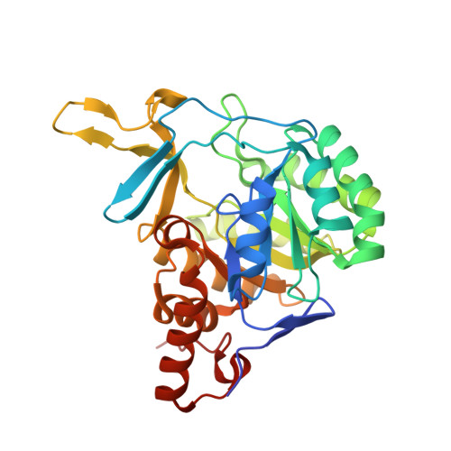
9VPX | pdb_00009vpx

Crystal structure of the C131A mutant of Trypanosoma brucei DHODH in FMN-oxidized, fumarate-bound form


Experimental Data Snapshot

  • Method: X-RAY DIFFRACTION
  • Resolution: 1.98 Å
  • R-Value Free: 
    0.215 (Depositor), 0.221 (DCC) 
  • R-Value Work: 
    0.179 (Depositor), 0.187 (DCC) 
  • R-Value Observed: 
    0.180 (Depositor) 

Starting Model: experimental
View more details

wwPDB Validation   3D Report Full Report


Ligand Structure Quality Assessment 


This is version 1.0 of the entry. See complete history


Literature

Structural Basis of Redox-Dependent Affinities of Dihydroorotate Dehydrogenase for Its Substrates and Products.

Tani, O.Kubota, T.Yamasaki, T.Hirokawa, T.Furukawa, K.Yamasaki, K.

(2025) J Mol Biology : 169544-169544

  • DOI: https://doi.org/10.1016/j.jmb.2025.169544
  • Primary Citation of Related Structures:  
    9VPR, 9VPS, 9VPT, 9VPU, 9VPV, 9VPW, 9VPX, 9VPY, 9VPZ, 9VQ0

  • PubMed Abstract: 

    Dihydroorotate dehydrogenase (DHODH), containing a cofactor, flavin mononucleotide (FMN), catalyzes the oxidation of dihydroorotate to orotate. In Trypanosoma brucei, the causative parasite of African trypanosomiasis, the DHODH enzyme (TbDHODH) couples this reaction with the reduction of fumarate to succinate. These reactions occur successively in the same catalytic site through a ping-pong bi-bi mechanism. FMN is reduced in the first step and re-oxidized in the second one. Here, we investigated how the redox state of FMN influences the binding of substrates and products to TbDHODH by NMR analyses of a catalytically impaired mutant. The affinities for the substrates, dihydroorotate and fumarate, were redox-dependent: dihydroorotate preferred the oxidized state, whereas fumarate preferred the reduced state. One of the products, succinate, exhibited no detectable binding to either of the redox states, suggesting its efficient product release. In contrast, the product of the first-half reaction, orotate, showed stronger binding to the oxidized state, suggesting that product inhibition may occur unless orotate is rapidly consumed in the subsequent biosynthetic step. The structural basis of these findings was elucidated by crystallography of the complexes with the above ligands in both redox states, along with quantitative analyses of the interactions using quantum chemical calculations. Upon reduction, the FMN isoalloxazine ring gains two additional hydrogens and becomes more bent at its center. These changes perturb the interactions among the ligands, flavin, and surrounding residues (notably Lys44, Asn68, and Asn195), primarily in the electrostatic term, which quantitatively explains the difference in binding affinities.


  • Organizational Affiliation
    • Biomedical Research Institute, National Institute of Advanced Industrial Science and Technology (AIST), 1-1-1 Higashi, Tsukuba 305-8566, Japan.

Macromolecules
Find similar proteins by:  (by identity cutoff)  |  3D Structure
Entity ID: 1
MoleculeChains Sequence LengthOrganismDetailsImage
Dihydroorotate dehydrogenase (fumarate)
A, B, C, D
317Trypanosoma brucei brucei TREU927Mutation(s): 2 
Gene Names: Tb927.5.3830
EC: 1.3.98.1
UniProt
Find proteins for Q57U83 (Trypanosoma brucei brucei (strain 927/4 GUTat10.1))
Explore Q57U83 
Go to UniProtKB:  Q57U83
Entity Groups  
Sequence Clusters30% Identity50% Identity70% Identity90% Identity95% Identity100% Identity
UniProt GroupQ57U83
Sequence Annotations
Expand
  • Reference Sequence
Small Molecules
Ligands 3 Unique
IDChains Name / Formula / InChI Key2D Diagram3D Interactions
FMN (Subject of Investigation/LOI)
Query on FMN

Download Ideal Coordinates CCD File 
E [auth A],
J [auth B],
N [auth C],
R [auth D]
FLAVIN MONONUCLEOTIDE
C17 H21 N4 O9 P
FVTCRASFADXXNN-SCRDCRAPSA-N
FUM (Subject of Investigation/LOI)
Query on FUM

Download Ideal Coordinates CCD File 
F [auth A],
K [auth B],
O [auth C],
S [auth D]
FUMARIC ACID
C4 H4 O4
VZCYOOQTPOCHFL-OWOJBTEDSA-N
MLI
Query on MLI

Download Ideal Coordinates CCD File 
G [auth A]
H [auth A]
I [auth A]
L [auth B]
M [auth B]
G [auth A],
H [auth A],
I [auth A],
L [auth B],
M [auth B],
P [auth C],
Q [auth C],
T [auth D]
MALONATE ION
C3 H2 O4
OFOBLEOULBTSOW-UHFFFAOYSA-L
Experimental Data & Validation

Experimental Data

  • Method: X-RAY DIFFRACTION
  • Resolution: 1.98 Å
  • R-Value Free:  0.215 (Depositor), 0.221 (DCC) 
  • R-Value Work:  0.179 (Depositor), 0.187 (DCC) 
  • R-Value Observed: 0.180 (Depositor) 
Space Group: P 21 21 2
Unit Cell:
Length ( Å )Angle ( ˚ )
a = 139.627α = 90
b = 146.529β = 90
c = 65.488γ = 90
Software Package:
Software NamePurpose
REFMACrefinement
PDB_EXTRACTdata extraction
HKL-2000data reduction
HKL-2000data scaling
PHASERphasing

Structure Validation

View Full Validation Report


Ligand Structure Quality Assessment 


Entry History & Funding Information

Deposition Data


Funding OrganizationLocationGrant Number
Not funded--

Revision History  (Full details and data files)

  • Version 1.0: 2025-11-26
    Type: Initial release