9VMJ | pdb_00009vmj

Crystal structure of TeGH116 from Thermosynechococcus elongatus


Experimental Data Snapshot

  • Method: X-RAY DIFFRACTION
  • Resolution: 2.20 Å
  • R-Value Free: 
    0.174 (Depositor), 0.184 (DCC) 
  • R-Value Work: 
    0.147 (Depositor), 0.160 (DCC) 

Starting Model: experimental
View more details

wwPDB Validation   3D Report Full Report


Ligand Structure Quality Assessment 


This is version 1.0 of the entry. See complete history


Literature

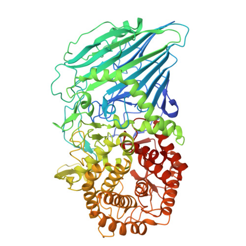
The structure of Thermosynechococcus elongatus glycoside hydrolase family 116 beta-glucosidase shows the role of the noncatalytic N-terminal domain in controlling substrate specificity.

Beagbandee, C.Charoenwattanasatien, R.Pengthaisong, S.Kurisu, G.Ketudat Cairns, J.R.

(2025) Int J Biol Macromol 332: 148566-148566

  • DOI: https://doi.org/10.1016/j.ijbiomac.2025.148566
  • Primary Citation of Related Structures:  
    9VMJ, 9VMK

  • PubMed Abstract: 

    β-Glucosidases play essential roles in nature as well as in industrial applications. Glycoside hydrolase family 116 (GH116) is a relatively sparsely characterized family of β-glucosidases. We describe the functional and structural characterization of Thermosynechococcus elongatus BP-1 TeGH116, the one β-glucosidase in this model cyanobacterium. TeGH116 was expressed in Escherichia coli and purified by heating, immobilized metal affinity chromatography, and size exclusion chromatography. TeGH116 hydrolyzes p-nitrophenyl β-d-glucopyranoside most rapidly at pH 5.5 and 55 °C, and other p-nitrophenyl glycosides 3% or less of this rate. TeGH116 also hydrolyzes natural phenolic glucosides and β-1,3- and β-1,4-linked gluco-oligosaccharides. Kinetic analysis suggests that TeGH116 binds three glucosyl residues in these oligosaccharides. TeGH116 is inhibited by glucose but is only weakly inhibited by δ-gluconolactone. The X-ray crystallographic structures of TeGH116 and its complex with glucose were similar to those of Thermoanaerobacterium xylanolyticum TxGH116. However, a long loop extending from the noncatalytic N-terminal domain helps form the mouth of the TeGH116 active site, making it narrower. TxGH116 was found to hydrolyze barley β-glucan, laminarin, and lichenan, but not carboxymethylcellulose, while TeGH116 hydrolyzed the same polysaccharides at relatively lower rates. Deletion of the loop in TeGH116 resulted in poor stability and low activity, but higher relative activity on laminarin compared to p-nitrophenyl β-d-glucoside. The narrowed active site and lower activity on large substrates highlight the contribution of N-terminal domain loops to the substrate specificity of GH116 enzymes.


  • Organizational Affiliation
    • Center for Biomolecular Structure, Function and Application, Suranaree University of Technology, 111 University Avenue, Muang, Nakhon Ratchasima, 30000, Thailand; School of Chemistry, Institute of Science, Suranaree University of Technology, 111 University Avenue, Muang, Nakhon Ratchasima, 30000, Thailand.

Macromolecules
Find similar proteins by:  (by identity cutoff)  |  3D Structure
Entity ID: 1
MoleculeChains Sequence LengthOrganismDetailsImage
Tlr0193 protein
A, B
809Thermosynechococcus vestitus BP-1Mutation(s): 0 
Gene Names: tlr0193
EC: 3.2.1.21
UniProt
Find proteins for Q8DMC7 (Thermosynechococcus vestitus (strain NIES-2133 / IAM M-273 / BP-1))
Explore Q8DMC7 
Go to UniProtKB:  Q8DMC7
Entity Groups  
Sequence Clusters30% Identity50% Identity70% Identity90% Identity95% Identity100% Identity
UniProt GroupQ8DMC7
Sequence Annotations
Expand
  • Reference Sequence
Small Molecules
Ligands 3 Unique
IDChains Name / Formula / InChI Key2D Diagram3D Interactions
TLA
Query on TLA

Download Ideal Coordinates CCD File 
G [auth A],
J [auth B]
L(+)-TARTARIC ACID
C4 H6 O6
FEWJPZIEWOKRBE-JCYAYHJZSA-N
GOL (Subject of Investigation/LOI)
Query on GOL

Download Ideal Coordinates CCD File 
C [auth A],
D [auth A],
E [auth A],
H [auth B]
GLYCEROL
C3 H8 O3
PEDCQBHIVMGVHV-UHFFFAOYSA-N
CA
Query on CA

Download Ideal Coordinates CCD File 
F [auth A],
I [auth B]
CALCIUM ION
Ca
BHPQYMZQTOCNFJ-UHFFFAOYSA-N
Experimental Data & Validation

Experimental Data

  • Method: X-RAY DIFFRACTION
  • Resolution: 2.20 Å
  • R-Value Free:  0.174 (Depositor), 0.184 (DCC) 
  • R-Value Work:  0.147 (Depositor), 0.160 (DCC) 
Space Group: C 1 2 1
Unit Cell:
Length ( Å )Angle ( ˚ )
a = 266.847α = 90
b = 55.299β = 129.724
c = 175.918γ = 90
Software Package:
Software NamePurpose
REFMACrefinement
HKL-2000data collection
autoPROCdata processing
MOLREPphasing

Structure Validation

View Full Validation Report


Ligand Structure Quality Assessment 


Entry History & Funding Information

Deposition Data


Funding OrganizationLocationGrant Number
Other governmentThailandSuranaree University of Technology
Other governmentThailandThailand Science Research and Innovation, and the National Science, Research and Innovation Fund (NSRF) under grant number 195669 (67A304000096)
Other governmentJapanOsaka University

Revision History  (Full details and data files)

  • Version 1.0: 2025-11-26
    Type: Initial release