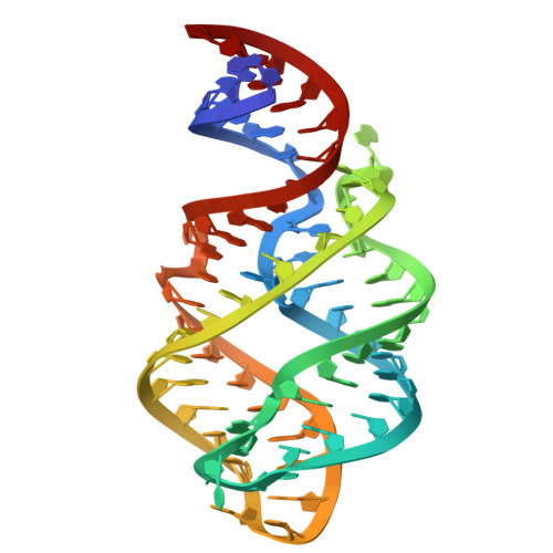
Ligand specificity and adaptability revealed by the first Guanine-II riboswitch tertiary structure.
Li, H., Shen, X., Xu, X., Tai, X., He, M., Zhang, J., Ren, A.(2025) Nucleic Acids Res 53
- PubMed: 40966503
- DOI: https://doi.org/10.1093/nar/gkaf884
- Primary Citation of Related Structures:
9LJN, 9LKC, 9LKE, 9LKF, 9LMC, 9V4U, 9V4V, 9V4X, 9V4Y, 9V4Z, 9V50, 9V51 - PubMed Abstract:
A comprehensive understanding of the fundamental principles governing RNA-small molecule interactions is crucial for advancing RNA-targeting therapeutics with small molecules. Riboswitches, a class of noncoding RNAs, regulate gene expression by direct interaction with small-molecule metabolites. In this work, we report an in-depth structure-based investigation of a newly identified riboswitch, Guanine-II, which, despite sharing a conserved scaffold with the Guanine-I riboswitch, exhibits strikingly distinct small molecule ligand-binding characteristics. Through a comprehensive structural analysis of the Guanine-II riboswitch bound to various guanine analogs, combined with comparative studies of other guanine riboswitch variants, including Guanine-I and Xanthine-II riboswitches, as well as isothermal titration calorimetry, we reveal local structural rearrangements that precisely modulate small-molecule ligand adaptability. We further demonstrate that subtle differences in the composition and peripheral architecture of the binding pocket are key determinants of ligand-binding specificity. Additionally, based on the similarity in ligand recognition patterns with the tetrahydrofolate-II riboswitch, we identified additional compounds that bind to the Guanine-II riboswitch through a structure-guided rational search, providing valuable structural insights for the discovery of small molecules targeting RNA.
- Department of Cardiology of The Second Affiliated Hospital and Life Sciences Institute and School of Medicine, Zhejiang University, Hangzhou 310058, China.
Organizational Affiliation:


