9R22 | pdb_00009r22

Cryo-EM structure of the light-driven proton pump PsPR in detergent micelle


Experimental Data Snapshot

  • Method: ELECTRON MICROSCOPY
  • Resolution: 2.48 Å
  • Aggregation State: PARTICLE 
  • Reconstruction Method: SINGLE PARTICLE 

wwPDB Validation   3D Report Full Report


This is version 1.0 of the entry. See complete history


Literature

Structural basis for no retinal binding in flotillin-associated rhodopsins.

Kovalev, K.Stetsenko, A.Trunk, F.Marin, E.Haro-Moreno, J.M.Lamm, G.H.U.Alekseev, A.Rodriguez-Valera, F.Schneider, T.R.Wachtveitl, J.Guskov, A.

(2025) Structure 

  • DOI: https://doi.org/10.1016/j.str.2025.06.006
  • Primary Citation of Related Structures:  
    9R21, 9R22, 9R23

  • PubMed Abstract: 

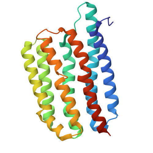
    Rhodopsins are light-sensitive membrane proteins capturing solar energy via a retinal cofactor covalently attached to a lysine residue. Several groups of rhodopsins lack the conserved lysine and showed no retinal binding. Recently, flotillin-associated rhodopsins (FArhodopsins or FARs) were identified and suggested to lack the retinal-binding pocket despite preserving the lysine residue in many members of the group. Here, we present cryoelectron microscopic (cryo-EM) structures of paralog FArhodopsin and proteorhodopsin from marine bacterium Pseudothioglobus, both forming pentamers similar to those of other microbial rhodopsins. We demonstrate no binding of retinal to the FArhodopsin despite preservation of the lysine residue and overall similarity of the protein fold and internal organization to those of the retinal-binding paralog. Mutational analysis confirmed that two amino acids, H84 and E120, prevent retinal binding within the FArhodopsin. Our work provides insights into the natural retinal loss in microbial rhodopsins and might contribute to the further understanding of FArhodopsins.


  • Organizational Affiliation

    European Molecular Biology Laboratory, 22607 Hamburg, Germany. Electronic address: kirill.kovalev@embl-hamburg.de.


Macromolecules
Find similar proteins by:  (by identity cutoff)  |  3D Structure
Entity ID: 1
MoleculeChains Sequence LengthOrganismDetailsImage
microbial rhodopsin
A, B, C, D, E
240Candidatus Pseudothioglobus sp.Mutation(s): 0 
Entity Groups  
Sequence Clusters30% Identity50% Identity70% Identity90% Identity95% Identity100% Identity
Sequence Annotations
Expand
  • Reference Sequence
Small Molecules
Ligands 3 Unique
IDChains Name / Formula / InChI Key2D Diagram3D Interactions
LMT
Query on LMT

Download Ideal Coordinates CCD File 
IA [auth D],
L [auth A],
LA [auth D],
NA [auth E],
U [auth B]
DODECYL-BETA-D-MALTOSIDE
C24 H46 O11
NLEBIOOXCVAHBD-QKMCSOCLSA-N
RET (Subject of Investigation/LOI)
Query on RET

Download Ideal Coordinates CCD File 
DA [auth C],
K [auth A],
KA [auth D],
SA [auth E],
T [auth B]
RETINAL
C20 H28 O
NCYCYZXNIZJOKI-OVSJKPMPSA-N
LFA
Query on LFA

Download Ideal Coordinates CCD File 
AA [auth C]
BA [auth C]
CA [auth C]
EA [auth D]
F [auth A]
AA [auth C],
BA [auth C],
CA [auth C],
EA [auth D],
F [auth A],
FA [auth D],
G [auth A],
GA [auth D],
H [auth A],
HA [auth D],
I [auth A],
J [auth A],
JA [auth D],
M [auth A],
MA [auth E],
N [auth B],
O [auth B],
OA [auth E],
P [auth B],
PA [auth E],
Q [auth B],
QA [auth E],
R [auth B],
RA [auth E],
S [auth B],
V [auth C],
W [auth C],
X [auth C],
Y [auth C],
Z [auth C]
EICOSANE
C20 H42
CBFCDTFDPHXCNY-UHFFFAOYSA-N
Experimental Data & Validation

Experimental Data

  • Method: ELECTRON MICROSCOPY
  • Resolution: 2.48 Å
  • Aggregation State: PARTICLE 
  • Reconstruction Method: SINGLE PARTICLE 

Structure Validation

View Full Validation Report


Entry History & Funding Information

Deposition Data


Funding OrganizationLocationGrant Number
Netherlands Organisation for Scientific Research (NWO)Netherlands184.034.014
Netherlands Organisation for Scientific Research (NWO)NetherlandsOCENW.KLEIN.141
EIPOD fellowship under Marie Sklodowska-Curie Actions COFUNDGermany847543

Revision History  (Full details and data files)

  • Version 1.0: 2025-07-23
    Type: Initial release