


A step forward for neuronal Nitric Oxide Synthase Inhibitors against Melanoma
Awasthi, A., Li, H., Hardy, C.D., Poulos, T.L., Silverman, R.B.To be published.
Experimental Data Snapshot
Starting Model: experimental
View more details
Entity ID: 1 | |||||
|---|---|---|---|---|---|
| Molecule | Chains | Sequence Length | Organism | Details | Image |
| Nitric oxide synthase, brain | 423 | Homo sapiens | Mutation(s): 2 Gene Names: NOS1 EC: 1.14.13.39 | 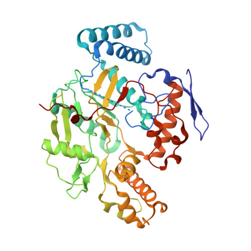 | |
UniProt & NIH Common Fund Data Resources | |||||
Find proteins for P29475 (Homo sapiens) Explore P29475 Go to UniProtKB: P29475 | |||||
PHAROS: P29475 GTEx: ENSG00000089250 | |||||
Entity Groups | |||||
| Sequence Clusters | 30% Identity50% Identity70% Identity90% Identity95% Identity100% Identity | ||||
| UniProt Group | P29475 | ||||
Sequence AnnotationsExpand | |||||
| |||||
| Ligands 5 Unique | |||||
|---|---|---|---|---|---|
| ID | Chains | Name / Formula / InChI Key | 2D Diagram | 3D Interactions | |
| HEM Query on HEM | E [auth A], K [auth B], O [auth C], U [auth D] | PROTOPORPHYRIN IX CONTAINING FE C34 H32 Fe N4 O4 KABFMIBPWCXCRK-RGGAHWMASA-L |  | ||
| A1BTY (Subject of Investigation/LOI) Query on A1BTY | G [auth A], M [auth B], Q [auth C], W [auth D] | N-[3-({[2-(4-{[(E)-(furan-2-yl)(imino)methyl]amino}phenyl)ethyl]amino}methyl)phenyl]furan-2-carboximidamide C25 H25 N5 O2 GNFGEHHGBYVPGG-UHFFFAOYSA-N |  | ||
| H4B Query on H4B | F [auth A], L [auth B], P [auth C], V [auth D] | 5,6,7,8-TETRAHYDROBIOPTERIN C9 H15 N5 O3 FNKQXYHWGSIFBK-RPDRRWSUSA-N |  | ||
| GOL Query on GOL | H [auth A] I [auth A] N [auth B] R [auth C] S [auth C] | GLYCEROL C3 H8 O3 PEDCQBHIVMGVHV-UHFFFAOYSA-N |  | ||
| ZN Query on ZN | J [auth B], T [auth C] | ZINC ION Zn PTFCDOFLOPIGGS-UHFFFAOYSA-N |  | ||
| Length ( Å ) | Angle ( ˚ ) |
|---|---|
| a = 118.482 | α = 90 |
| b = 52.261 | β = 90 |
| c = 165.052 | γ = 90 |
| Software Name | Purpose |
|---|---|
| PHENIX | refinement |
| Aimless | data scaling |
| XDS | data reduction |
| REFMAC | phasing |
| Funding Organization | Location | Grant Number |
|---|---|---|
| National Institutes of Health/National Institute of General Medical Sciences (NIH/NIGMS) | United States | GM131920 |


