


The Structural Basis of NMN Synthesis Catalyzed by NadV from Haemophilus ducreyi
Lin, T., Zhengjuan, W., Jia, Y.To be published.
Experimental Data Snapshot
Starting Model: in silico
View more details
Entity ID: 1 | |||||
|---|---|---|---|---|---|
| Molecule | Chains | Sequence Length | Organism | Details | Image |
| Nicotinamide phosphoribosyltransferase | 495 | [Haemophilus] ducreyi | Mutation(s): 0 Gene Names: nadV, PNAD10009, RZ57_05785 EC: 2.4.2.12 | 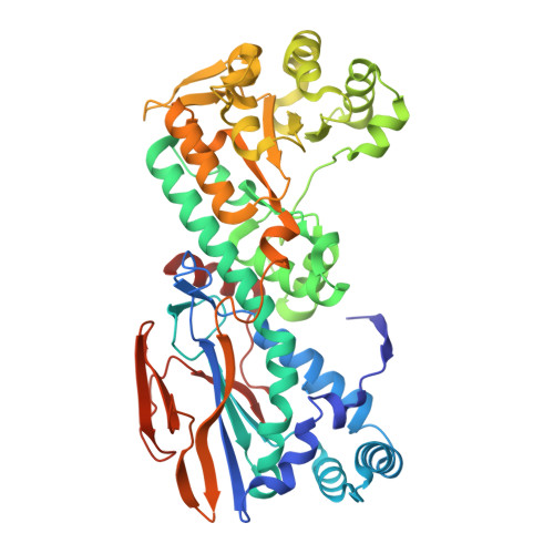 | |
UniProt | |||||
Find proteins for G1U9V7 (Haemophilus ducreyi) Explore G1U9V7 Go to UniProtKB: G1U9V7 | |||||
Entity Groups | |||||
| Sequence Clusters | 30% Identity50% Identity70% Identity90% Identity95% Identity100% Identity | ||||
| UniProt Group | G1U9V7 | ||||
Sequence AnnotationsExpand | |||||
| |||||
| Ligands 1 Unique | |||||
|---|---|---|---|---|---|
| ID | Chains | Name / Formula / InChI Key | 2D Diagram | 3D Interactions | |
| NMN (Subject of Investigation/LOI) Query on NMN | C [auth A], D [auth B] | BETA-NICOTINAMIDE RIBOSE MONOPHOSPHATE C11 H16 N2 O8 P DAYLJWODMCOQEW-TURQNECASA-O |  | ||
| Length ( Å ) | Angle ( ˚ ) |
|---|---|
| a = 158.52 | α = 90 |
| b = 158.52 | β = 90 |
| c = 130.4 | γ = 120 |
| Software Name | Purpose |
|---|---|
| PHENIX | refinement |
| XDS | data reduction |
| Aimless | data scaling |
| MOLREP | phasing |
| Funding Organization | Location | Grant Number |
|---|---|---|
| National Natural Science Foundation of China (NSFC) | China | -- |


