9JTZ | pdb_00009jtz

Crystal structure of Nir2 C-terminal domain in complex with phosphatidic acid


Experimental Data Snapshot

  • Method: X-RAY DIFFRACTION
  • Resolution: 2.15 Å
  • R-Value Free: 
    0.258 (Depositor), 0.227 (DCC) 
  • R-Value Work: 
    0.203 (Depositor), 0.222 (DCC) 
  • R-Value Observed: 
    0.205 (Depositor) 

Starting Model: experimental
View more details

wwPDB Validation   3D Report Full Report


Ligand Structure Quality Assessment 


This is version 1.1 of the entry. See complete history


Literature

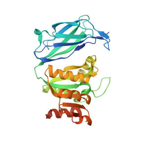
Nir2 crystal structures reveal a phosphatidic acid-sensing mechanism at ER-PM contact sites.

Kim, D.Lee, S.Jun, Y.Lee, C.

(2025) Proc Natl Acad Sci U S A 122: e2516849122-e2516849122

  • DOI: https://doi.org/10.1073/pnas.2516849122
  • Primary Citation of Related Structures:  
    9JTZ, 9JUI, 9JYX, 9JZ1, 9JZP

  • PubMed Abstract: 

    Agonist-induced activation of phosphoinositide-specific phospholipase C (PLC) converts phosphatidylinositol 4,5-bisphosphate [PI(4,5)P 2 ] to diacylglycerol (DAG) at the inner leaflet of the plasma membrane (PM). DAG can be enzymatically transformed into phosphatidic acid (PA) and accumulated at the PM. PYK2 N-terminal domain-interacting receptor 2 (Nir2) mediates the formation of ER-PM membrane contact sites (MCSs) by specifically recognizing PA at the PM and directly interacting with ER membrane protein vesicle-associated membrane protein-associated proteins (VAPs). The N-terminal phosphatidylinositol transfer protein domain of Nir2 facilitates PI/PA exchange at ER-PM MCSs to maintain PI and PA levels. Here, we reveal the mechanisms by which Nir2 senses phosphatidic acid (PA) and associates with membranes, based on three crystal structures of its C-terminal Lipin/Ned1/Smp2 (LNS2) domain bound to PA, the diphenylalanine [FF]-containing acidic tract (FFAT) motif complexed with vesicle-associated membrane protein-associated protein B/C (VAPB), and the Asp-Asp-His-Asp (DDHD) domain. The C-terminal LNS2 domain of Nir2 directly interacts with the phosphate in the headgroup of PA via hydrogen bonds involving S1025, T1065, K1103, and K1126. Formation of a salt bridge between E355 in Nir2 and R55 in VAPB is essential for Nir2 FFAT-VAPB interaction. The central DDHD domain of Nir2 forms a twofold symmetric dimer, and this self-association contributes to stable and tight membrane association. These findings reveal how Nir2-mediated ER-PM MCS formation maintains continued PI(4,5)P 2 -dependent PLC signaling.


  • Organizational Affiliation
    • Department of Life Sciences, Korea University, Seoul 02841, South Korea.

Macromolecules
Find similar proteins by:  (by identity cutoff)  |  3D Structure
Entity ID: 1
MoleculeChains Sequence LengthOrganismDetailsImage
Membrane-associated phosphatidylinositol transfer protein 1
A, B, C
284Mus musculusMutation(s): 0 
Gene Names: Pitpnm1Dres9Mpt1Nir2Pitpnm
UniProt & NIH Common Fund Data Resources
Find proteins for O35954 (Mus musculus)
Explore O35954 
Go to UniProtKB:  O35954
IMPC:  MGI:1197524
Entity Groups  
Sequence Clusters30% Identity50% Identity70% Identity90% Identity95% Identity100% Identity
UniProt GroupO35954
Sequence Annotations
Expand
  • Reference Sequence
Experimental Data & Validation

Experimental Data

  • Method: X-RAY DIFFRACTION
  • Resolution: 2.15 Å
  • R-Value Free:  0.258 (Depositor), 0.227 (DCC) 
  • R-Value Work:  0.203 (Depositor), 0.222 (DCC) 
  • R-Value Observed: 0.205 (Depositor) 
Space Group: P 21 21 21
Unit Cell:
Length ( Å )Angle ( ˚ )
a = 69.941α = 90
b = 108.764β = 90
c = 120.535γ = 90
Software Package:
Software NamePurpose
PHENIXrefinement
HKL-2000data scaling
HKL-2000data reduction
PHENIXphasing

Structure Validation

View Full Validation Report


Ligand Structure Quality Assessment 


Entry History & Funding Information

Deposition Data


Funding OrganizationLocationGrant Number
National Research Foundation (NRF, Korea)Korea, Republic OfNRF-2021M3A9G8022417
National Research Foundation (NRF, Korea)Korea, Republic OfNRF-2021R1A2C2009550

Revision History  (Full details and data files)

  • Version 1.0: 2025-10-15
    Type: Initial release
  • Version 1.1: 2025-11-05
    Changes: Database references