Elucidating the structure and assembly mechanism of actinoporin pores in complex membrane environments.
Arranz, R., Santiago, C., Masiulis, S., Rivera-de-Torre, E., Palacios-Ortega, J., Carlero, D., Heras-Marquez, D., Gavilanes, J.G., Arias-Palomo, E., Martinez-Del-Pozo, A., Garcia-Linares, S., Martin-Benito, J.(2025) Sci Adv 11: eadv0683-eadv0683
- PubMed: 40991702
- DOI: https://doi.org/10.1126/sciadv.adv0683
- Primary Citation of Related Structures:
9GJ8, 9GKI, 9GKL, 9GKO, 9GKP - PubMed Abstract:
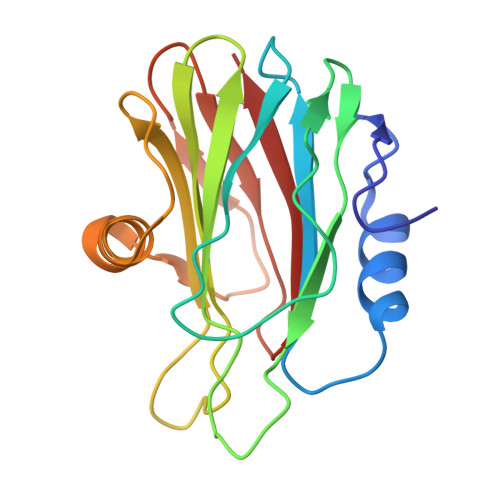
Pore-forming proteins exemplify the transformative potential of biological molecules. Produced as soluble monomers, they assemble into multimeric membrane-inserted complexes in response to specific membrane environments. Actinoporins, a class of pore-forming proteins from sea anemones, target membranes to kill cells. Here, we report cryogenic electron microscopy structures of two actinoporins, fragaceatoxin C and sticholysin II, reconstituted in lipid membranes. The structures reveal an ordered arrangement of dozens of lipid molecules that form an integral part of the pore architecture. We also captured distinct oligomeric intermediates, arc-shaped assemblies with monomers in transitional conformations, representing key snapshots along the pore formation pathway. These data provide direct structural evidence for a stepwise mechanism in which monomers sequentially bind the membrane and undergo conformational changes that drive pore assembly and membrane disruption. Our findings reveal how these proteins reshape membranes and offer mechanistic insights into their cytolytic activity. This work broadens our understanding of pore-forming proteins, which are gaining increasing relevance in diverse biotechnological applications.
- Departamento de Estructura de Macromoléculas, Centro Nacional de Biotecnología, Consejo Superior de Investigaciones Científicas, Calle Darwin 3, 28049 Madrid, Spain.
Organizational Affiliation: