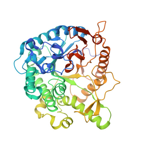
Neutron crystallography of the covalent intermediate of beta-glucosidase reveals remodeling of the catalytic center.
Yano, N., Kashima, T., Arakawa, H., Lin, C.C., Ishiwata, A., Namiki, H., Takaya, N., Tanaka, K., Kusaka, K., Fushinobu, S.(2025) Proc Natl Acad Sci U S A 122
- PubMed: 41166426
- DOI: https://doi.org/10.1073/pnas.2502828122
- Primary Citation of Related Structures:
9LPH, 9LPI, 9LPV, 9LPX, 9LPY - PubMed Abstract:
Anomer-retaining glycoside hydrolases (GHs) generally catalyze a double displacement reaction via a covalent intermediate. However, neutron crystallography of glycoside ligand-bound states has not been performed. In this study, we investigated β-glucosidase Td2F2 from GH family 1 as a model enzyme for anomer-retaining GHs. We determined joint X-ray/neutron structures of Td2F2 in ligand-free form, covalent intermediate with a 2-deoxy-2-fluoro glucoside (2F-Glc) inhibitor, and glucose product complex using hydrogen/deuterium-exchanged crystals at room temperature, with neutron diffraction resolutions of 1.80-1.70 Å. Extensive hydrogen bonds recognizing the hydroxy groups of 2F-Glc were identified, along with the positions of deuterium atoms. The acid/base catalyst residue Glu166 was anchored by a hydrogen bond network pivoted by Asn293. Tyr295 forms a hydrogen bond with the catalytic nucleophile residue Glu352 in the ligand-free and glucose complex forms, while the active center undergoes significant reorganization, including side chain displacements of Glu352 and Tyr295, as well as the incorporation of a water molecule. An alternative conformation of Tyr295 was observed in the 2F-Glc structure at room temperature, suggesting its role in positioning the nucleophilic water during the deglycosylation step. Steady-state and pre-steady-state kinetic analyses of Y295F mutant supported the functional involvement of Tyr295 in both glycosylation and deglycosylation steps. The tyrosine hydrogen bonded to the nucleophile is also conserved in many other anomer-retaining GH families, underscoring its importance in catalysis. Based on the deuterium/hydrogen positions determined from neutron structures, we proposed a detailed reaction mechanism for Td2F2.
- Structural Biology Division, Japan Synchrotron Radiation Research Institute, Sayo-gun, Hyogo 679-5198, Japan.
Organizational Affiliation:


