


Crystal structure of Saccharomyces cerevisiae cross-linked Sfh1 (K197C, F233C) mutant in complex with phosphatidylethanolamine
Green, S.M., Laganowsky, A., Krieger, I., Singh, P.K., Sacchettini, J., Igumenova, T.I., Bankaitis, V.A.To be published.
Experimental Data Snapshot
Starting Model: experimental
View more details
Entity ID: 1 | |||||
|---|---|---|---|---|---|
| Molecule | Chains | Sequence Length | Organism | Details | Image |
| CRAL-TRIO domain-containing protein YKL091C | 310 | Saccharomyces cerevisiae | Mutation(s): 3 Gene Names: YKL091C | 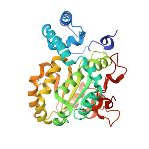 | |
UniProt | |||||
Find proteins for P33324 (Saccharomyces cerevisiae (strain ATCC 204508 / S288c)) Explore P33324 Go to UniProtKB: P33324 | |||||
Entity Groups | |||||
| Sequence Clusters | 30% Identity50% Identity70% Identity90% Identity95% Identity100% Identity | ||||
| UniProt Group | P33324 | ||||
Sequence AnnotationsExpand | |||||
| |||||
| Ligands 3 Unique | |||||
|---|---|---|---|---|---|
| ID | Chains | Name / Formula / InChI Key | 2D Diagram | 3D Interactions | |
| PTY (Subject of Investigation/LOI) Query on PTY | C [auth A], H [auth B] | PHOSPHATIDYLETHANOLAMINE C40 H80 N O8 P NJGIRBISCGPRPF-KXQOOQHDSA-N |  | ||
| SO4 (Subject of Investigation/LOI) Query on SO4 | D [auth A] E [auth A] F [auth A] G [auth A] K [auth B] | SULFATE ION O4 S QAOWNCQODCNURD-UHFFFAOYSA-L |  | ||
| GOL (Subject of Investigation/LOI) Query on GOL | I [auth B], J [auth B] | GLYCEROL C3 H8 O3 PEDCQBHIVMGVHV-UHFFFAOYSA-N |  | ||
| Length ( Å ) | Angle ( ˚ ) |
|---|---|
| a = 49.43 | α = 90 |
| b = 72.01 | β = 93.95 |
| c = 114.68 | γ = 90 |
| Software Name | Purpose |
|---|---|
| PHENIX | refinement |
| XDS | data reduction |
| XDS | data scaling |
| MOLREP | phasing |
| Funding Organization | Location | Grant Number |
|---|---|---|
| Welch Foundation | United States | A-2194-20240404 |
| Robert A. Welch Foundation | United States | BE-0017 |
| National Institutes of Health/National Institute of General Medical Sciences (NIH/NIGMS) | United States | NIH R35 GM131804 |
| National Institutes of Health/National Institute of General Medical Sciences (NIH/NIGMS) | United States | NIH RO1 GM108998 |


