7UY0 | pdb_00007uy0

Crystal structure of human Fgr tyrosine kinase in complex with A-419259


Experimental Data Snapshot

  • Method: X-RAY DIFFRACTION
  • Resolution: 2.55 Å
  • R-Value Free: 
    0.223 (Depositor), 0.220 (DCC) 
  • R-Value Work: 
    0.186 (Depositor), 0.190 (DCC) 
  • R-Value Observed: 
    0.188 (Depositor) 

Starting Model: experimental
View more details

wwPDB Validation   3D Report Full Report


Ligand Structure Quality Assessment 


This is version 1.3 of the entry. See complete history


Literature

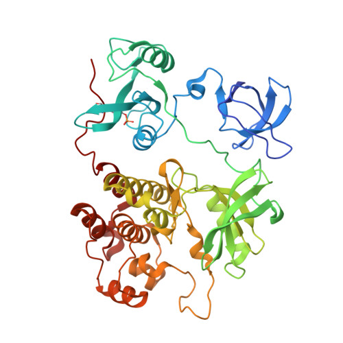
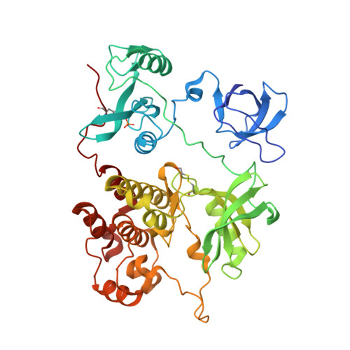
ATP-site inhibitors induce unique conformations of the acute myeloid leukemia-associated Src-family kinase, Fgr.

Du, S.Alvarado, J.J.Wales, T.E.Moroco, J.A.Engen, J.R.Smithgall, T.E.

(2022) Structure 30: 1508-1517.e3

  • DOI: https://doi.org/10.1016/j.str.2022.08.008
  • Primary Citation of Related Structures:  
    7UY0, 7UY3

  • PubMed Abstract: 

    The Src-family kinase Fgr is expressed primarily in myeloid hematopoietic cells and contributes to myeloid leukemia. Here, we present X-ray crystal structures of Fgr bound to the ATP-site inhibitors A-419259 and TL02-59, which show promise as anti-leukemic agents. A-419259 induces a closed Fgr conformation, with the SH3 and SH2 domains engaging the SH2-kinase linker and C-terminal tail, respectively. In the Fgr:A-419259 complex, the activation loop of one monomer inserts into the active site of the other, providing a snapshot of trans-autophosphorylation. By contrast, TL02-59 binding induced SH2 domain displacement from the C-terminal tail and SH3 domain release from the linker. Solution studies using HDX MS were consistent with the crystal structures, with A-419259 reducing and TL02-59 enhancing solvent exposure of the SH3 domain. These structures demonstrate that allosteric connections between the kinase and regulatory domains of Src-family kinases are regulated by the ligand bound to the active site.


  • Organizational Affiliation
    • Department of Microbiology and Molecular Genetics, University of Pittsburgh School of Medicine, Bridgeside Point II, Suite 523, 450 Technology Drive, Pittsburgh, PA 15219, USA.

Macromolecules
Find similar proteins by:  (by identity cutoff)  |  3D Structure
Entity ID: 1
MoleculeChains Sequence LengthOrganismDetailsImage
Tyrosine-protein kinase Fgr456Homo sapiensMutation(s): 4 
Gene Names: FGRSRC2
EC: 2.7.10.2
UniProt & NIH Common Fund Data Resources
Find proteins for P09769 (Homo sapiens)
Explore P09769 
Go to UniProtKB:  P09769
PHAROS:  P09769
GTEx:  ENSG00000000938 
Entity Groups  
Sequence Clusters30% Identity50% Identity70% Identity90% Identity95% Identity100% Identity
UniProt GroupP09769
Sequence Annotations
Expand
  • Reference Sequence
Find similar proteins by:  (by identity cutoff)  |  3D Structure
Entity ID: 2
MoleculeChains Sequence LengthOrganismDetailsImage
Tyrosine-protein kinase Fgr456Homo sapiensMutation(s): 4 
Gene Names: FGRSRC2
EC: 2.7.10.2
UniProt & NIH Common Fund Data Resources
Find proteins for P09769 (Homo sapiens)
Explore P09769 
Go to UniProtKB:  P09769
PHAROS:  P09769
GTEx:  ENSG00000000938 
Entity Groups  
Sequence Clusters30% Identity50% Identity70% Identity90% Identity95% Identity100% Identity
UniProt GroupP09769
Sequence Annotations
Expand
  • Reference Sequence
Small Molecules
Ligands 4 Unique
IDChains Name / Formula / InChI Key2D Diagram3D Interactions
VSE (Subject of Investigation/LOI)
Query on VSE

Download Ideal Coordinates CCD File 
E [auth A],
H [auth B]
7-[trans-4-(4-methylpiperazin-1-yl)cyclohexyl]-5-(4-phenoxyphenyl)-7H-pyrrolo[2,3-d]pyrimidin-4-amine
C29 H34 N6 O
FDVSOQRNTAPCHB-YHBQERECSA-N
PO4
Query on PO4

Download Ideal Coordinates CCD File 
L [auth B],
M [auth B]
PHOSPHATE ION
O4 P
NBIIXXVUZAFLBC-UHFFFAOYSA-K
GOL
Query on GOL

Download Ideal Coordinates CCD File 
C [auth A],
D [auth A],
G [auth B]
GLYCEROL
C3 H8 O3
PEDCQBHIVMGVHV-UHFFFAOYSA-N
CL
Query on CL

Download Ideal Coordinates CCD File 
F [auth A],
I [auth B],
J [auth B],
K [auth B]
CHLORIDE ION
Cl
VEXZGXHMUGYJMC-UHFFFAOYSA-M
Modified Residues  2 Unique
IDChains TypeFormula2D DiagramParent
PTR
Query on PTR
A
L-PEPTIDE LINKINGC9 H12 N O6 PTYR
CME
Query on CME
B
L-PEPTIDE LINKINGC5 H11 N O3 S2CYS
Experimental Data & Validation

Experimental Data

  • Method: X-RAY DIFFRACTION
  • Resolution: 2.55 Å
  • R-Value Free:  0.223 (Depositor), 0.220 (DCC) 
  • R-Value Work:  0.186 (Depositor), 0.190 (DCC) 
  • R-Value Observed: 0.188 (Depositor) 
Space Group: P 21 21 21
Unit Cell:
Length ( Å )Angle ( ˚ )
a = 49.43α = 90
b = 112.09β = 90
c = 213.73γ = 90
Software Package:
Software NamePurpose
PHENIXrefinement
XDSdata reduction
XSCALEdata scaling
PDB_EXTRACTdata extraction
PHASERphasing

Structure Validation

View Full Validation Report


Ligand Structure Quality Assessment 


Entry History & Funding Information

Deposition Data


Funding OrganizationLocationGrant Number
National Institutes of Health/National Cancer Institute (NIH/NCI)United StatesCA233576

Revision History  (Full details and data files)

  • Version 1.0: 2022-12-28
    Type: Initial release
  • Version 1.1: 2023-10-25
    Changes: Data collection, Refinement description
  • Version 1.2: 2023-11-15
    Changes: Data collection
  • Version 1.3: 2024-11-13
    Changes: Structure summary