2ATF | pdb_00002atf

X-RAY STRUCTURE OF cysteine dioxygenase type I FROM MUS MUSCULUS MM.241056


Experimental Data Snapshot

  • Method: X-RAY DIFFRACTION
  • Resolution: 1.75 Å
  • R-Value Free: 
    0.216 (Depositor), 0.223 (DCC) 
  • R-Value Work: 
    0.179 (Depositor), 0.190 (DCC) 
  • R-Value Observed: 
    0.181 (Depositor) 

wwPDB Validation   3D Report Full Report


This is version 1.4 of the entry. See complete history


Literature

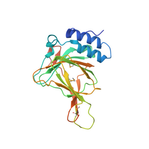
Structure and mechanism of mouse cysteine dioxygenase.

McCoy, J.G.Bailey, L.J.Bitto, E.Bingman, C.A.Aceti, D.J.Fox, B.G.Phillips, G.N.

(2006) Proc Natl Acad Sci U S A 103: 3084-3089

  • DOI: https://doi.org/10.1073/pnas.0509262103

  • PubMed Abstract: 

    Cysteine dioxygenase (CDO) catalyzes the oxidation of l-cysteine to cysteine sulfinic acid. Deficiencies in this enzyme have been linked to autoimmune diseases and neurological disorders. The x-ray crystal structure of CDO from Mus musculus was solved to a nominal resolution of 1.75 Angstroms. The sequence is 91% identical to that of a human homolog. The structure reveals that CDO adopts the typical beta-barrel fold of the cupin superfamily. The NE2 atoms of His-86, -88, and -140 provide the metal binding site. The structure further revealed a covalent linkage between the side chains of Cys-93 and Tyr-157, the cysteine of which is conserved only in eukaryotic proteins. Metal analysis showed that the recombinant enzyme contained a mixture of iron, nickel, and zinc, with increased iron content associated with increased catalytic activity. Details of the predicted active site are used to present and discuss a plausible mechanism of action for the enzyme.


  • Organizational Affiliation

    Center for Eukaryotic Structural Genomics and Department of Biochemistry, University of Wisconsin, Madison, 53706-1544, USA.


Macromolecules
Find similar proteins by:  (by identity cutoff)  |  3D Structure
Entity ID: 1
MoleculeChains Sequence LengthOrganismDetailsImage
Cysteine dioxygenase type I200Mus musculusMutation(s): 3 
Gene Names: Cdo1
EC: 1.13.11.20
UniProt
Find proteins for P60334 (Mus musculus)
Explore P60334 
Go to UniProtKB:  P60334
Entity Groups  
Sequence Clusters30% Identity50% Identity70% Identity90% Identity95% Identity100% Identity
UniProt GroupP60334
Sequence Annotations
Expand
  • Reference Sequence
Small Molecules
Modified Residues  1 Unique
IDChains TypeFormula2D DiagramParent
MSE
Query on MSE
A
L-PEPTIDE LINKINGC5 H11 N O2 SeMET
Experimental Data & Validation

Experimental Data

  • Method: X-RAY DIFFRACTION
  • Resolution: 1.75 Å
  • R-Value Free:  0.216 (Depositor), 0.223 (DCC) 
  • R-Value Work:  0.179 (Depositor), 0.190 (DCC) 
  • R-Value Observed: 0.181 (Depositor) 
Space Group: P 43 21 2
Unit Cell:
Length ( Å )Angle ( ˚ )
a = 57.55α = 90
b = 57.55β = 90
c = 122.076γ = 90
Software Package:
Software NamePurpose
DENZOdata reduction
SCALEPACKdata scaling
SHARPphasing
DMphasing
REFMACrefinement
PDB_EXTRACTdata extraction

Structure Validation

View Full Validation Report


Entry History 

Revision History  (Full details and data files)

  • Version 1.0: 2005-10-18
    Type: Initial release
  • Version 1.1: 2007-10-16
    Changes: Version format compliance
  • Version 1.2: 2011-07-13
    Changes: Version format compliance
  • Version 1.3: 2017-10-11
    Changes: Refinement description
  • Version 1.4: 2024-10-30
    Changes: Data collection, Database references, Derived calculations, Structure summary